journal menu


issue contents
March 2009 issue
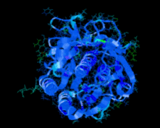
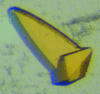
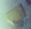
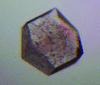
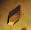
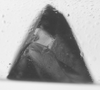
Cover illustration: High-resolution structure of proteinase K cocrystallized with digalacturonic acid (Larson et al., p. 192).
structural communications


The crystal structure of proteinase K cocrystallized with digalacturonic acid and HEPES was solved at 1.32 Å resolution. The disaccharide was rotationally disordered about a twofold axis, but nonetheless contributed to intermolecular lattice interactions.
PDB reference: proteinase K–digalacturonic acid complex, 3dyb, r3dybsf
The crystal structure of a hyperthermophilic D-tagatose 3-epimerase-related protein with a unique active-site architecture was determined.
PDB reference: D-tagatose 3-epimerase-related protein, 2zvr, r2zvrsf
Open  access
access
The structure of the MarR-family regulator NMB1585 from N. meningitidis has been solved using data extending to 2.1 Å resolution.
PDB reference: NMB1585, 3g3z, r3g3zsf
crystallization communications
A 2.5 Å resolution data set was collected from a crystal of a soluble chimeric form of NADPH-cytochrome P450 reductase (CPR) produced using a fusion gene composed of the yeast FMN and the human FAD domains. The chimeric protein was crystallized in a modified conformation compared with the previously solved structures.
Canavalia boliviana lectin (Cbol) was purified using a Sephadex G-50 column and crystallized in the presence of X-Man by hanging-drop vapour diffusion at 293 K. After optimization, crystals suitable for diffraction were obtained using 0.1 M HEPES pH 7.5 and 3.0 M sodium formate.
The aminoacylhistidine dipeptidase encoded by V. alginolyticus pepD has been overexpressed and crystallized.
The thermophilic cellulase FnCel5A from F. nodosum Rt17-B1 has been overexpressed in E. coli, purified and crystallized. Diffraction data were collected to 2.4 Å resolution for the native enzyme and to 1.7 Å resolution for the selenomethionyl derivative.
Recombinant nucleotide-binding domain of E. coli Mg-ATPase was expressed, purified and used to produce crystals that diffracted to 1.53 Å resolution.
Two members of the class C family of bacterial nonspecific acid phosphatases have been cloned, expressed, purified and crystallized. One of the crystal forms exhibited epitaxial twinning.
The LADI-III diffractometer at the Institut Laue–Langevin has been used to carry out the first neutron crystallographic study of a DNA oligonucleotide in the A conformation. The crystal size was 0.06 mm3, the smallest ever used successfully for a study of this type. The results provide evidence of unexpected base protonation and illustrate the opportunities that now exist for nucleic acid crystallography using both hydrogenated and perdeuterated oligonucleotides.
The site-specific unglycosylated mutant of metabotropic glutamate receptor 3 extracellular domain has been overexpressed, purified, and crystallized. A complete data set has been collected to 2.35 Å resolution.
Open access
access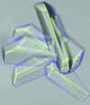
Three fusion proteins were generated in order to resolve the atomic structure of the CFA/I fimbriae of enterotoxigenic E. coli. CfaEB is a fusion of the minor and major CFA/I subunits, while CfaBB and CfaBBB are tandem fusions of two and three repeats, respectively, of the major subunit. Each protein was crystallized and the crystal structures of each of these fusions were determined successively by the molecular-replacement method using the CfaE crystal structure as an initial phasing model.
Two C-terminal fragments of the RNA helicase Hera have been crytallized in three crystal forms, one of which was phased by MAD using a single selenium site.
Dihydrodipicolinate synthase (DHDPS) catalyzes an important step in lysine biosynthesis. Here, the crystallization and preliminary diffraction analysis to 1.2 Å resolution of DHDPS from C. botulinum in the presence of its substrate pyruvate is reported.
The cofactor-independent monooxygenase SnoaB from the nogalamycin-biosynthesis pathway and a designed selenomethionine-substituted double mutant were crystallized in space group P21212. The native enzyme diffracted to a resolution of 2.4 Å.
CmlS from S. venezuelae is a flavin-dependent halogenase that is involved in the biosynthesis of the widely used antibiotic chloramphenicol. Here, the crystallization of CmlS and analysis of the initial diffraction data are reported.
A putative two-domain-type laccase from a metagenome was crystallized using the sitting-drop vapour-diffusion method. Diffraction data were collected to a resolution of 1.7 Å.
The Z-DNA-binding domain of a PKR-like kinase (PKZ) from goldfish was crystallized in complex with d(TCGCGCG)2. The crystals belonged to space group C2 and diffracted to 1.7 Å resolution.
Two cuticle-degrading proteases Ver112 and PL646 were purified from the nematophagous fungi L. psalliotae and P. lilacinus, respectively. The protease Ver112 and a complex between PL646 and a tetrapeptide inhibitor were crystallized. Diffraction data were collected to 1.65 and 2.2 Å resolution, respectively.
A cohesin-like module from the hyperthermophilic archaeon A. fulgidus was cloned, expressed, purified and crystallized. X-ray diffraction data were collected to 1.82 Å resolution.
Here, the C-terminal domain of the human voltage-gated proton channel Hv1 (C-Hv1) was overexpressed in Escherichia coli, purified and crystallized using the hanging-drop vapour-diffusion method.
In order to further illustrate the catalytic mechanism of arginine decarboxylase by determining the three-dimensional structure of the enzyme the speA gene was amplified from B. subtilis genomic DNA and cloned. The enzyme was expressed in Escherichia coli and purified to homogeneity by nickel-chelation chromatography followed by size-exclusion chromatography. High-quality crystals were obtained using the hanging-drop vapour-diffusion method at 298 K.
An RNA-binding region of human HuR bound to an 11-base RNA fragment has been crystallized. The crystals diffracted to a resolution of 1.8 Å and belonged to space group P212121.
α-Enolase from human liver (hENO1) was expressed as a soluble protein and purified by affinity column chromatography and gel filtration. Crystals were obtained by the hanging-drop vapour-diffusion method and diffracted to 2.5 Å resolution.
A C-terminal fragment of VERNALIZATION1 from A. thaliana, a DNA-binding protein required for the acceleration of flowering in response to prolonged cold treatment, was crystallized. X-ray diffraction data were collected to a resolution of 2.1 Å.
The formation, crystallization and preliminary X-ray diffraction analysis of the tumour necrosis factor α (TNF)–tumour necrosis factor receptor type 2 (TNFR2) complex are described. The initial electron-density map, which was calculated using only the phases of refined TNF trimer structures, could detect the main chains and side chains of TNFR2 around the TNF trimer.
Wild-type and mutant recombinant human transforming growth factor β-induced protein (TGFBIp) were cloned, purified and crystallized. Preliminary X-ray crystallography data were obtained from wild-type TGFBIp.
The cloning and expression of recombinant PilZXAC1133, a protein belonging to the PilZ superfamily, are described. PilZ proteins are associated with the control of several complex behaviours in bacteria and in some cases have been shown to bind c-diGMP. PilZXAC1133 containing selenomethionine produced crystals that diffracted to 1.85 Å resolution.
Recombinant selenomethionine-labelled LipL32, the major surface protein of pathogenic Leptospira, has been purified and crystallized. Data sets from two crystals were collected, one of which diffracted to 2.25 Å resolution.
Est-Y29, a novel oligomeric class C β-lactamase from a metagenomic library, was crystallized in space group I41 and diffraction data were collected to 1.49 Å resolution.
Preliminary studies were carried out to purify and crystallize the sample from cat (Felis silvestris catus), a low oxygen-affinity haemoglobin in different crystal forms.
A 2.0 Å resolution neutron diffraction data set has been collected from a D2O-soaked γ-chymotrypsin crystal at low pH on the Institute Laue–Langevin LADI-III beamline.
![[publBio]](/logos/publbio.gif)