journal menu


issue contents
January 2022 issue
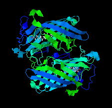
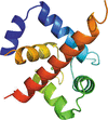
Cover illustration: DynF from the dynemicin-biosynthesis pathway of Micromonospora chersina. Access to the biosynthetic gene cluster of dynemicin, an enediyne natural product, has enabled the in vitro study of gene products within the cluster to decipher their roles in assembling this unique molecule. The crystal structure of one of these gene products, DynF, is reported by Kosgei et al. [(2022), Acta Cryst. F78, 1–7].
research communications


Open  access
access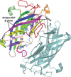
The crystal structure of DynF was determined to a resolution of 1.50 Å, revealing a dimeric eight-stranded β-barrel structure with palmitic acid bound in the interior.
PDB reference: DynF from Micromonospora chersina, 6ubl
Open access
access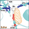
Human mitochondrial manganese superoxide dismutase (MnSOD) is a major player in alleviating oxidative stress within the mitochondria, which is a characteristic of a wide range of human diseases. Here, the methods leading to the structural visualization of the evasive peroxo complex of MnSOD by integrating cryotrapping and neutron protein crystallography are described.
The X-ray crystal structure of a human cardiac muscle troponin C/troponin I chimera has been determined in two different crystal forms and shows a conformation of the complex that differs from that previously observed by NMR. In contrast to previous models, the troponin I helix runs in a parallel direction relative to the troponin C groove and completely blocks the calcium desensitizer binding site of the troponin C–troponin I interface.
Open access
access
The high-resolution structure of a putative short-chain reductase from the commercially important bacterium Paraburkholderia xenovorans is reported. P. xenovorans degrades organic wastes such as polychlorinated biphenyls.
PDB reference: putative short-chain reductase, 5jc8
Open access
access
Crystal structures of FolM alternative dihydrofolate reductase 1 from Brucella suis and Brucella canis reveal prototypical NADPH-dependent short-chain reductases with structural similarity to protozoan pteridine reductases that are potential drug targets.
The crystallization of the SALM3–PTPσ synaptic adhesion complex and the initial low-resolution structure solution by molecular replacement is presented. This independently verifies the architecture of the ligand complex to be similar to the SALM5–PTPδ crystal structure and complements our previous small-angle X-ray scattering studies.

![[publBio]](/logos/publbio.gif)