journal menu


issue contents
September 2005 issue
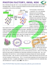
Cover illustration: Composite image showing various aspects of high-pressure XRD experiment: panoramic diamond anvil cell (DAC) used to generate pressure, sample in a DAC, composed of five magnesiowustite single crystals and a ruby sphere (see Jacobsen et al., pages 577-583), polychromatic (bottom left, image courtesy of P. Dera) and monochromatic diffraction images from samples of different crystallinity ranging from single crystal to amorphous (see Prakapenka et al., pages 560-565), experimental set-up for synchrotron polychromatic microdiffraction (see Ice et al., pages 608-617) and example of crystal structure of a post-perovskite high-pressure phase (courtesy of C. T. Prewitt).
facility information


SXD at Mbar pressures


Free 
Open access
access
The use of data from poor-quality single crystals, and from combined powder/single-crystal studies, to determine crystal structures at high pressure is reviewed.
Combining perforated diamond anvil cells with focusing optics at a third-generation synchrotron X-ray source allows collection of scattering data of sufficient quality to perform real-space modeling of the pair distribution function obtained from nano-crystalline materials to 10 GPa.
Grain size and grain boundaries are important parameters that should be taken into account in high-pressure diffraction studies. The effects of pressure and temperature on crystal growth in the diamond-anvil cell have been studied using various starting materials.
Structure changes induced by lattice–electron interactions under high pressure have been investigated using a diamond anvil pressure cell.
Single-crystal samples of wüstite-Fe0.93O and magnesiowüstite-(Mg0.73Fe0.27)O were hydrostatically compressed in a diamond-anvil cell to 51 GPa. Equation-of-state parameters for the high-pressure rhombohedral phase of Fe0.93O were determined.
Phase decomposition is considered for structure solutions in binary systems at high pressure and high temperature.

Download citation


Download citation
Open access
access
The fully ordered high-pressure crystal structure of cyclopentanol (C5H10O) has been solved using single-crystal X-ray diffraction techniques on station 9.8 at the SRS Daresbury Laboratory. The crystal structure is characterized by the formation of molecular chains, denoted C (4) in graph set notation, with the molecules adopting a pseudo fourfold arrangement around the central core of hydrogen bonds.
Download citation
Download citation
Open access
access
The structure of hexagonal L-cystine consists of pairs of hydrogen-bonded layers connected by disulfide bridges. The conformation about the bridges changes on compression to 3.7 GPa as the C—S—S—C units act like springs; other changes can be understood by examination of the distribution of voids in the ambient pressure structure.
Applications of various modifications of polychromatic single-crystal microdiffraction to experiments at ultrahigh pressure are discussed.
Open access
access
Recent developments in solid-state imaging technologies have provided a framework for faster, lower noise and larger format systems than previously available. Advances which have not found widespread use in instrumentation for X-ray diffraction include highly parallel readout amplifiers, hybrid imaging technologies and improved CMOS imagers. These advances are discussed and simulations are presented of selected devices incorporated into a new generation of instrumentation for Laue diffraction applications.

Strategies for reducing preferred orientation and strain in powder samples for high-pressure synchrotron X-ray diffraction in the diamond-anvil cell are presented.
The possibility of using X-ray diffraction to precisely monitor crystal structure at the extremes of pressure and temperature is explored. A summary of the advantages of using various X-ray sources for this work and an outline of the necessary experimental layout is given.
X-ray emission spectroscopy and X-ray diffraction have been used in conjunction with the double-sided laser-heated diamond cell technique for studying local electronic spin states of ferrous iron in magnesiowüstite-(Mg0.75,Fe0.25)O and its crystal structure under high pressures and temperatures in situ. The new method can be used to study 3d transition metals in oxides and silicates under conditions relevant to deep-Earth and planetary interiors.
Instrumentation for high-pressure research with the diamond anvil cell at GSECARS is described in terms of capabilities and characteristics. Future prospects are discussed with regard to these facilities for high-pressure research.

A new high-pressure facility for diffraction and spectroscopy using diamond anvil high-pressure cells has been developed at the Advanced Light Source. Details of the mechanics and performance of the beamline and endstation are given.
A new high-flux high-pressure XRD beamline is now in operation at the ESRF.
research papers

A new precise synchrotron-ring synchronized X-ray beam shutter has been built and tested.
A new Compton spectrometer, constructed at beamline ID15B of the ESRF, is described. This spectrometer is based on a novel idea, the dispersion compensation.
A versatile X-ray absorption cell is presented giving the possibility of investigating model catalysts at atmospheric pressure and powder catalysts under plug-flow conditions. As an example, Au/SiO2 model and powder catalysts are examined.
The design of a new cell for in situ and operando X-ray absorption experiments is reported. The cell is successfully tested for in situ characterization of cobalt silica-supported Fischer-Tropsch catalysts and operando studies of methanol oxidation over molybdenum catalysts.
The time-dependent deformation tensor, having dilational and shear components, has been determined by time-resolved triple-crystal X-ray diffractometry. The observed dynamic deformation in a GaAs wafer irradiated by ultrashort laser pulses, i.e. pulsed photothermal deformation, was followed by the appearance of a flexural standing wave.
The use of a portable video telescope to obtain X-ray excited optical luminescence spectra is described, including examples of light emission from luminescent organic molecules.
A phase problem and spectrum recovery from the modulus of an interferogram are discussed.
An analyzer-based X-ray phase contrast imaging system with an asymmetrical crystal set-up has been mounted for quantitative and qualitative studies at the SYRMEP beamline of Elettra. The technique is shown to be powerful enough to see details in ox brain tissue that are not detected using conventional synchrotron radiography.
new commercial products
Free
current events
Free