

 menu
|
|
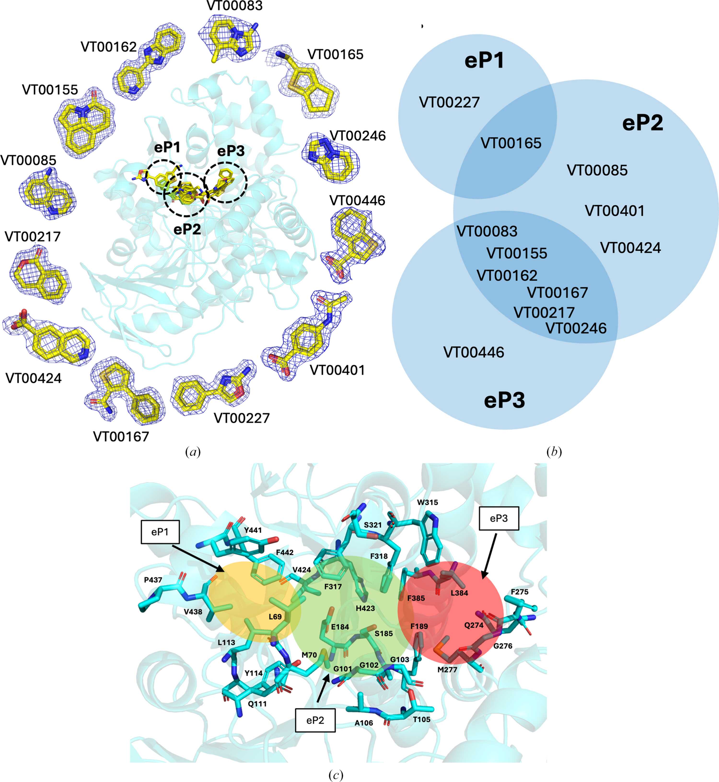
Figure 4
Fragment-based active-site mapping/probing identifies potential pockets for plastic binding to esterase AXPUR5. (a) Superimposed structures of fragments for esterase with each ligand demonstrated separately. The 2mFo − DFc standard map at a 1.0σ contour level for esterase fragments is shown as a blue mesh. (b) The overlap of fragments that bound to eP1 (two hits), eP2 (ten hits) and eP3 (seven hits) is shown in circles with relative sizes corresponding to the number of hits. (c) A close-up view of the fragment-binding pockets with surrounding residues highlighted and the pockets shown in shaded colours. |
 | STRUCTURAL BIOLOGY |
ISSN: 2059-7983
Open  access