

 menu
|
|
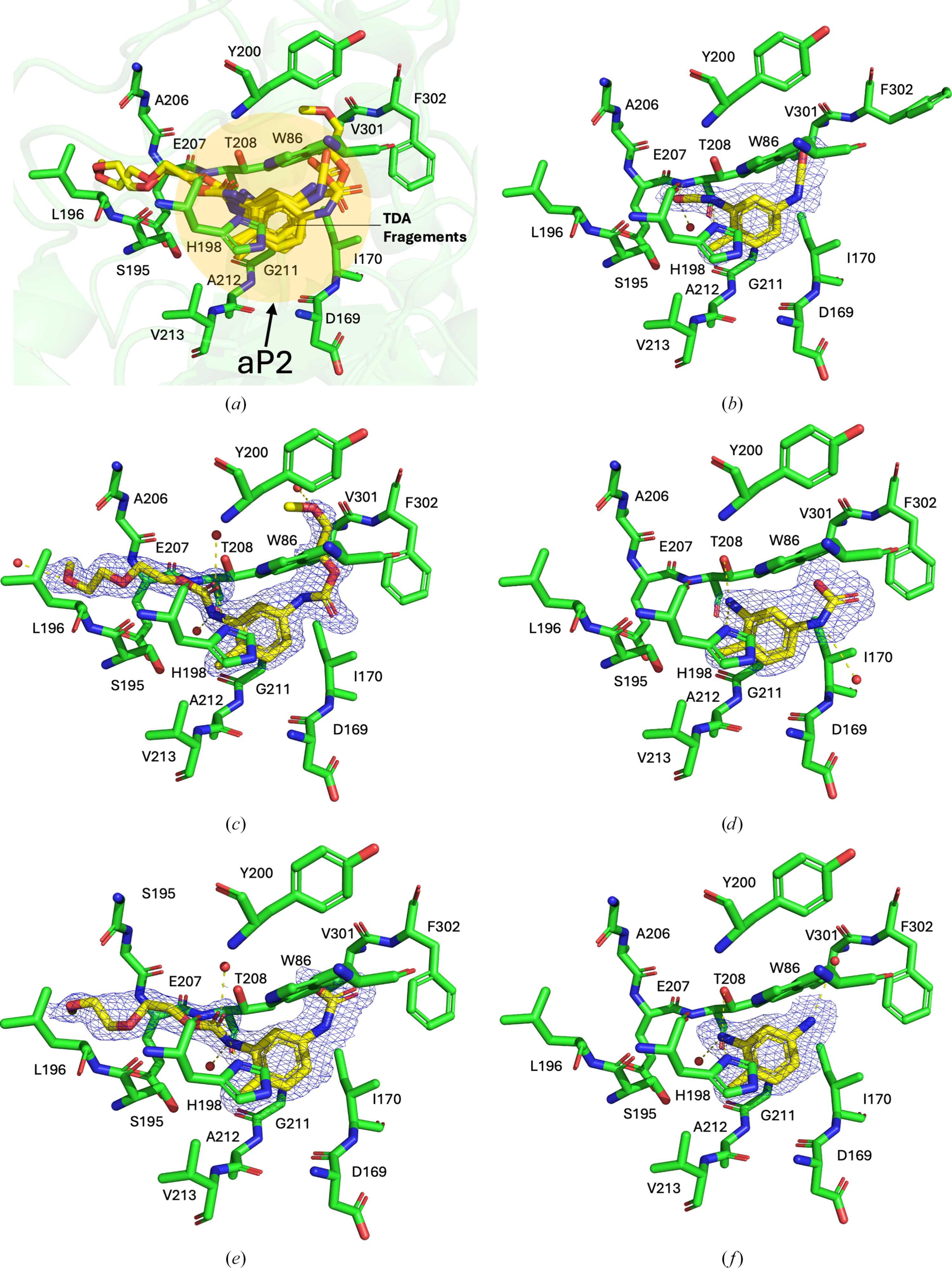
Figure 5
Validation of subpockets as plastic fragment-binding sites in amidase U11. (a) Superimposed structures of bound fragments in aP2 and surrounding residues in the pocket are shown. (b) 2,4-TDI (PDB entry 30qw). (c) DUE-2,4-TDA (PDB entry 30hf). (d) 2,4-TDA (carbamic acid; PDB entry 30hg). (e) MUE-2,4-TDA (carbamic acid; PDB entry 30nq). (f) 2,4-TDA (amine; PDB entry 30rg). 2mFo − DFc maps at a 1.0σ contour level are shown as a blue mesh for the ligand along with surrounding residues. Hydrogen bonds are shown as dashed yellow lines and water molecules are depicted as red spheres. |
 | STRUCTURAL BIOLOGY |
ISSN: 2059-7983
Open  access