

 menu
|
|
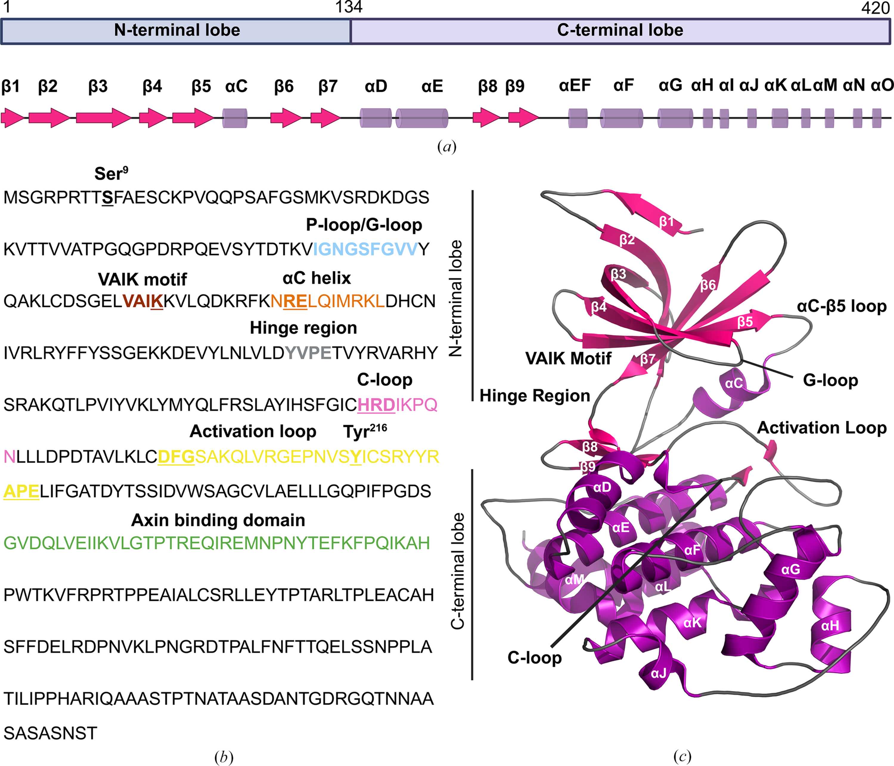
Figure 1
Structural domains and regulatory features of GSK3β. (a) Graphical representation of GSK3β domains (top) and secondary-structure elements (bottom). (b) Human GSK3β amino-acid sequence (residues 1–420), with key structural and regulatory regions highlighted: the phosphate-binding loop (P-loop), also known as the glycine rich-loop (G-loop; residues 62–70, sky blue), the VAIK motif (residues 82–85; maroon), the αC-helix (residues 95–104, orange), the hinge region (residues 134–137, gray), the catalytic loop (C-loop, 182–186, light pink), the HRD motif (residues 179–181, light pink), the activation loop (residues 200–226, yellow) and the axin-binding domain (ABD, residues 262–299, green). The regulatory residues Ser9, Lys85, Arg96, Glu97, His179, Arg180, Asp181, Asp200, Phe201, Gly202, Tyr216, Ala224, Pro225 and Glu226 are underlined. (c) A three-dimensional structure of GSK3β showing various regulatory regions (PDB entry 9huk). The β-strands, α-helices and loops are shown in hot pink, purple and gray, respectively. (a) was created in https://BioRender.com. |
 | STRUCTURAL BIOLOGY |
ISSN: 2059-7983
Open  access