

 menu
|
|
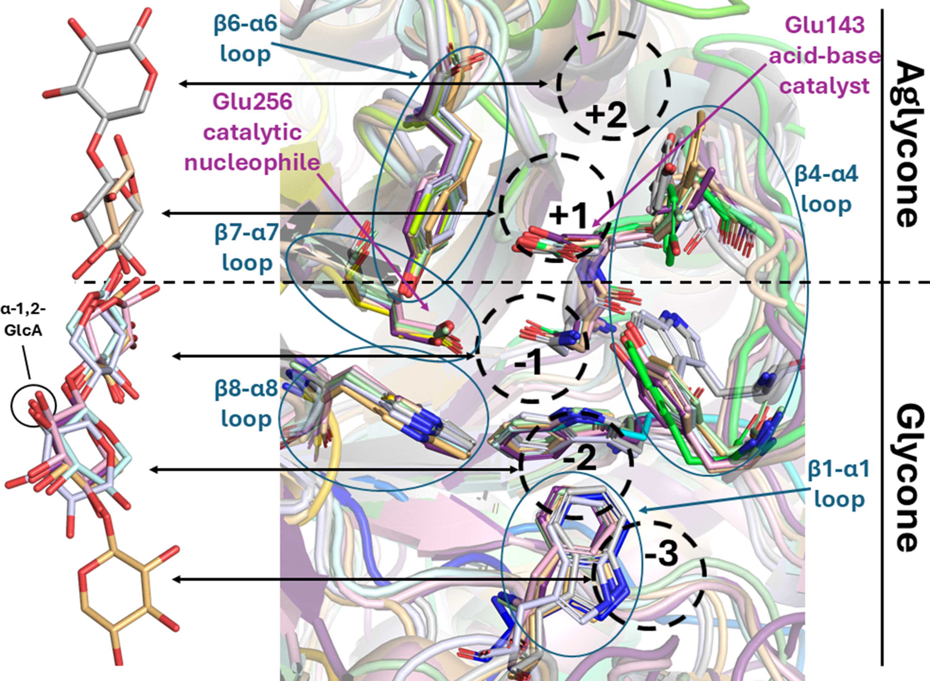
Figure 5
Amino-acid conservation in the XBC. Pair fitting alignment reveals the closeness between related GH30 subfamilies. The overlaid sugar ligands resulting from the alignment are shifted to the left side for clarity and subsites are indicated as well as the catalytic center and five loop regions. Pair fitting of AchXyn30A (in rainbow) with two GH30_7 dual-function GX/XBH enzymes [TcXyn30B (PDB entry 6krn) in pale cyan and TtXyn30A (PDB entry 7o0e) in light pink], a GH30_7 enzyme with REX activity and a GH30_7 EX with −2 subsite affinity [TcXyn30A (PDB entry 8idq) in wheat and TcXyn30C (PDB entry 6m5z) in pale green], three GH30_8 bacterial GXs [EcXyn30A (PDB entry 2y24) in light orange, BsXyn30C (PDB entry 3kl5) in blue white and CtXyn30A (PDB entry 5a6m) in gray] and lastly a GH30_10 XBH [AcXbh30A (PDB entry 7n6o) in light blue]. The glycone region ligands obtained from the listed GX GH30_8 enzymes each contained a GlcA substituted α-1,2 on the −2 subsite xylose. This appendage has been removed for clarity and its location is noted in the figure. |
 | STRUCTURAL BIOLOGY |
ISSN: 2059-7983
