
 journal menu


short communications
 | JOURNAL OF SYNCHROTRON RADIATION |
Development of an (X, eX) spectrometer for measuring the energies of the scattered photon and recoil electron

aDepartment of Synchrotron Radiation Science, The Graduate University for Advanced Studies, 1-1 Oho, Tsukuba, Ibaraki 305-0801, Japan, bPhoton Factory, KEK, 1-1 Oho, Tsukuba, Ibaraki 305-0801, Japan, and cDepartment of Electronic Engineering, Faculty of Engineering, Gunma University, 1-5-1 Tenjincho, Kiryu, Gunma 376-8515, Japan
*Correspondence e-mail: [email protected]
The design and performance of a new spectrometer for coincidence measurements between the Compton scattered photon and the recoil electron are described. Coincidence measurements give direct information on the three-dimensional electron momentum density (EMD) of condensed matter. The present spectrometer measures energy spectra of both the photon and the electron. The energy spectrum of electrons is measured by a time-of-flight method using single-bunch operation at the Photon Factory Accumulator Ring (PF-AR). The energy resolution obtained for the recoil electron is 190 eV, which is better than that of the photon detector, so that a momentum resolution of the three-dimensional EMD of 0.3 can be achieved.
Keywords: Compton scattering; coincident technique; time of flight; single bunch.
1. Introduction
The coincidence measurement between the Compton scattered photon and the recoil electron (X, eX) provides direct information on the three-dimensional electron momentum density (EMD). The feasibility of such measurements by detecting the directions of the Compton scattered photon and the recoil electron, and the energy of the photon has been reported by several authors (Rollason et al., 1989![[Rollason, A. J., Bell, F. & Schneider, J. R. (1989). Nucl. Instrum. Methods A, 281, 147-155.]](../../../../../../logos/arrows/s_arr.gif "Rollason, A. J., Bell, F. & Schneider, J. R. (1989). Nucl. Instrum. Methods A, 281, 147-155.") ; Bell et al., 1990; Tschentscher et al., 1993; Kurp et al., 1996). The momentum space resolution obtained is limited by the energy resolution of the photon detector and it is difficult to obtain a momentum resolution better than 0.5 a.u. (atomic units). The energy width of the EMD for free and/or valence electrons in condensed matter is about 1 a.u. Therefore, it is necessary to obtain a much better momentum space resolution in order to study Fermiology and/or electron-binding energies. We have developed a new spectrometer which enables us to measure not only the above three components, i.e. the directions of the Compton scattered photon and the recoil electron, and the energy of the photon, but also the energy of the recoil electron by means of a time-of-flight method (TOF). The TOF measurement has given us the energy spectrum of the recoil electron with an energy resolution better than that of a photon detector, allowing us to determine the three-dimensional EMD for graphite with a momentum resolution of 0.3 a.u.
; Bell et al., 1990; Tschentscher et al., 1993; Kurp et al., 1996). The momentum space resolution obtained is limited by the energy resolution of the photon detector and it is difficult to obtain a momentum resolution better than 0.5 a.u. (atomic units). The energy width of the EMD for free and/or valence electrons in condensed matter is about 1 a.u. Therefore, it is necessary to obtain a much better momentum space resolution in order to study Fermiology and/or electron-binding energies. We have developed a new spectrometer which enables us to measure not only the above three components, i.e. the directions of the Compton scattered photon and the recoil electron, and the energy of the photon, but also the energy of the recoil electron by means of a time-of-flight method (TOF). The TOF measurement has given us the energy spectrum of the recoil electron with an energy resolution better than that of a photon detector, allowing us to determine the three-dimensional EMD for graphite with a momentum resolution of 0.3 a.u.
2. Kinetics
Fig. 1 shows a diagram of the Compton scattering process. An incident photon with the four-component vector k1 = (k1, iω1) makes an inelastic collision with an electron p1 = (p1, iE1). The photon is scattered to angle θ with k2 = (k2, iω2) and the electron is recoiled with p2 = (p2, iE2). In an impulse approximation, the fourfold cross section is given by equation (1) using natural units (i.e. ![[\hbar]](teximages/he3146fi1.gif) = m = c = 1) (Rollason et al., 1989)
= m = c = 1) (Rollason et al., 1989)

where α is the fine-structure constant and X is the function as shown in (2)
![[\eqalign{X& = (R/R^{\prime}) +(R^{\prime}/R) -\sin^2 \theta\cr R& = -{\bf p}_1 \cdot {\bf k}_1 = -{\bf p}_2\cdot {\bf k}_2\cr R^{\prime}& = -{\bf p}_1\cdot {\bf k}_2 = -{\bf p}_2 \cdot {\bf k}_1.\cr}\eqno(2)]](teximages/he3146fd2.gif)
In the coincidence measurement case, the cross section is proportional to the electron momentum density itself
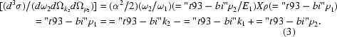
For coincidence events, we have the following energy conservation law
![[\hbar\omega_1 = \hbar\omega_2 +T +E_b \eqno(4)]](teximages/he3146fd4.gif)
where T is the recoil electron energy and Eb is the electron-bonding energy. The high-resolution measurement of T gives us ρ(p1) at high momentum resolution via the combination of equations (3) and (4).
![[Figure 1]](he3146fig1thm.gif) | Figure 1 Momentum diagram of the Compton scattering processes. |
3. Design of the spectrometer
Fig. 2 shows the plan and side views of the spectrometer. The incident X-rays with energy ω = 65 keV impinge on the sample, which is placed at the centre of a sample chamber. The chamber is evacuated by turbo-molecular pumps and its inside is covered by permalloy to avoid the effect of magnetic fields. Samples are placed on a translation stage in order to adjust the position. Recoil electrons are detected by a multi-channel-plate (MCP) electron detector (Hamamatsu F2223-21SX) which is placed at a distance of L = 2.00 m from the sample under an angle of φ = 12.5°. The diameter of the MCP is 27 mm. The T of the recoil electron is determined by the time of flight t, as T = (L/t)2/2m. The energy resolution ΔT/T is equal to 2Δt/t where Δt is the overall time resolution determined by both the intrinsic bunch width of the synchrotron radiation and the time resolution of the electronics. A PIN diode with thickness 200 µm is placed upstream of the sample and serves as a timing trigger. Compton scattered photons are detected by a segmented Ge solid-state detector (SSD) that covers a scattering angle θ = 148–154°. The diameter of each element is 8 mm and the space between them is 14 mm. According to equation (3), the different scattering angles of the SSD k2 provide us with different three-dimensional EMDs with a corresponding lateral momentum py simultaneously. The SSD elements are placed at py = −0.81, −0.36, 0.09, 0.55, 1.10 a.u. corresponding to a, b, c, d, e in Fig. 2 (top right). In order to measure three-dimensional EMDs for py > 1.0, the chamber is designed to be rotated around a pivot in its centre. The resolution of py is 0.5 a.u. and is defined by the solid angles accepted by the photon and electron detectors.
![[Figure 2]](he3146fig2thm.gif) | Figure 2 A side and plan view of the spectrometer and the arrangement of the SSD elements. |
Fig. 3 shows the electronic circuit of the spectrometer. All signals from the SSD and the MCP are collected by the multi-parameter analyser (FAST/Comtech MPA-Win) in a list mode. We can obtain two kinds of coincidence data: the so-called electron branch (photon branch) which represents a measurement of the energy spectrum of recoil electrons (scattered photons) in coincidence with scattered photons (recoil electrons). We use eight inputs of the MPA in order to measure simultaneously seven outputs of the segmented SSDs that are placed at different scattering angles.
![[Figure 3]](he3146fig3thm.gif) | Figure 3 A block diagram of the electronic circuit of the (X, eX) spectrometer. |
4. Performance (results and discussion)
The spectrometer was installed at the Photon Factory Accumulator Ring (PF-AR) NE1 beamline (KEK). The PF-AR, whose energy and stored current were 6.5 GeV and 40 mA, respectively, was operated in single-bunch mode. The time interval between two pulses was 1.28 µs. The white X-rays from an elliptical multipole wiggler, which was installed in the NE1 beamline, were monochromated by a doubly bent crystal monochromator (Kawata et al., 1998). The energy of the monochromated X-rays was chosen to be 65 keV with an energy resolution ΔE/E ≃ 10−3. The beam size at the sample was about 0.5 mm (vertical) × 3 mm (horizontal). The sample was a graphite foil with a thickness of 20 nm. The count rate of the MCP (SSD) from the graphite foil was about 15 counts s−1 (50 counts s−1 per element). A coincidence gate time of 1.6 µs was used and the coincidence count rate was approximately 3 counts min−1. This yielded a signal-to-noise ratio of 60.
Figs. 4(a) and 4(b) show TOF spectra for the non-coincidence and the coincidence measurements, respectively. There are three peaks in Fig. 4(a). These are located at 6.7, 14.5 and ∼32 ns and correspond to scattered photons, photoelectrons (65 keV) and recoil electrons (∼12.5 keV). The peak of the recoil electrons corresponds to the double-integrated EMD. Thus, it has a long tail caused by core-electron contributions. The full width at half-maximum (FWHM) of the photon peak (230 ps) corresponds to the overall time resolution of the system. The main contribution to the peak width is the bunch length of 200 ps. Then, the energy resolution for the recoil electrons caused by the above time resolution is 190 eV, corresponding to 0.30 a.u. momentum resolution along pz. In the coincidence measurement, Fig. 4(b), there is only the recoil electron peak and the profile gives us direct information about the three-dimensional EMD. Similarly, Figs. 4(c) and 4(d) show energy spectra of the scattered photons in the non-coincidence and coincidence measurements, respectively. The peak in Fig. 4(d) corresponds to the three-dimensional EMD in the photon branch. The energy resolution of the SSD is 480 eV at 65 keV which corresponds to 0.70 a.u. momentum resolution along pz. Figs. 4(e) and 4(f) show the three-dimensional EMDs for graphite derived from the electron and photon branches. The three-dimensional EMD in the electron branch looks like a flat top, but that in the photon branch looks like a Gaussian peak. This discrepancy is due to the difference in resolution (0.3 and 0.7 a.u.). The solid lines in Figs. 4(e) and 4(f) represent theoretical calculations of the three-dimensional EMD given by Kheifets & Vos (1995) and convoluted with the respective resolutions. The agreement with the experimental data is very good.
![[Figure 4]](he3146fig4thm.gif) | Figure 4 Experimental results of non-coincidence and coincidence measurements. Each spectrum shows the TOF profile of an electron in (a) non-coincidence and (b) coincidence cases and likewise the energy profile of a photon in (c) non-coincidence and (d) coincidence cases. (e) and (f) show the three-dimensional EMDs obtained in the electron and photon branches, respectively. |
Fig. 5 shows the results of ρ(0, py, pz) for graphite by using an array of photon detectors. The three-dimensional EMDs for different py (−0.9–2.2 a.u.) were obtained from two data sets of φ = 12.5 and 16.8°. In this way, the three-dimensional EMD of a graphite foil could be measured with Δpz = 0.3 a.u. and Δpy = 0.5 a.u. resolutions in momentum space.
![[Figure 5]](he3146fig5thm.gif) | Figure 5 The electron momentum density of graphite: ρ(0, py, pz). |
Acknowledgements
The authors gratefully acknowledge Professor F. Bell, University of Munich, for providing us with the graphite sample of 20 nm thickness. The measurements were performed with the approval of the Photon Factory Advisory Committee, Proposal No. 94-G363.
References
 Bell, F., Rollason, A. J., Schneider, J. R. & Drube, W. (1990). Phys. Rev. B, 41, 4887–4890. CrossRef Web of Science
Kawata, H., Sato, M., Higashi, Y. & Yamaoka, H. (1998). J. Synchrotron Rad. 5, 673–675. Web of Science CrossRef CAS IUCr Journals
Kheifets, A. S. & Vos, M. (1995). J. Phys. Condens. Matter, 7, 3895–3904. CrossRef CAS Web of Science
Kurp, F. F., Tschentscher, Th., Schulte-Schrepping, H., Schneider, J. R. & Bell, F. (1996). Europhys. Lett. 35, 61–66. CrossRef CAS Web of Science
Rollason, A. J., Bell, F. & Schneider, J. R. (1989). Nucl. Instrum. Methods A, 281, 147–155. CrossRef Web of Science
Tschentscher, Th., Schneider, J. R. & Bell, F. (1993). Phys. Rev. B, 48, 16965–16973. CrossRef CAS Web of Science
© International Union of Crystallography. Prior permission is not required to reproduce short quotations, tables and figures from this article, provided the original authors and source are cited. For more information, click here.
| JOURNAL OF SYNCHROTRON RADIATION |


