
 journal menu


issue contents
 | STRUCTURAL BIOLOGY |
ISSN: 2059-7983
Image-processing methods for electron microscopy of biological specimens
Edited by Carlos Oscar Sorzano, Alberto Bartesaghi and Amit Singer
This virtual issue collects together articles from the 2024 call for papers on image-processing methods for electron microscopy of biological specimens.
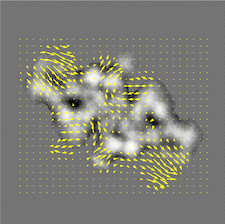
Cover illustration: The ManifoldEM method for conformational heterogeneity analysis in single-particle cryo-EM has been made more accessible. An optical flow visualization for thyroglobulin projection directions is shown.


Free 

The focused issue on Image-processing methods for electron microscopy of biological specimens is introduced. The virtual issue is available at https://journals.iucr.org/special_issues/2025/imageprocessing.

Open access
access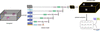
TomoCPT: a generalizable model for 3D particle detection and localization in cryo-electron tomograms
TomoCPT is a generalizable transformer-based 3D particle-picking tool for cryo-tomographic data.
The ManifoldEM method for conformational heterogeneity analysis in single-particle cryo-EM was first presented nearly a decade ago; however, its accessibility to a wide audience has been limited. Here, we describe a step-by-step breakdown accompanied by a modern Python implementation with increased usability, enhanced performance and improved accessibility to future feature additions.
Open access
access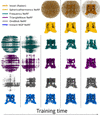
Cryo-EM density-map inference, with fixed pose and contrast transfer function, using a multi-resolution hash-encoding framework called instant-NGP, is described, together with its extension to heterogeneity inference by bending space with a per-image vector field.
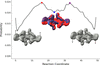
In the context of cryo-EM reconstruction, we provide a theoretical analysis showing that the condition number of the optimization problem is particularly large at high resolution, leading to slow convergence of gradient-based methods such as stochastic gradient descent. We then propose a method to overcome this issue by computing a preconditioner using Hutchinson's estimator, which results in improved convergence speed, as evidenced by numerical experiments.
We present CryoLike, a user-friendly Python workflow for calculating the image-to-structure likelihood in cryo-electron microscopy.


