

 journal menu


issue contents
April 2002 issue
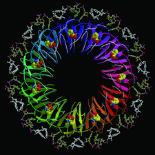
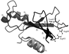
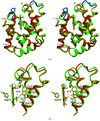
Cover illustration: B. subtilis trp RNA-binding attenuation protein (TRAP) complexed with an RNA containing 11 GAG triplets. TRAP is shown as a ribbon with each of the 11 subunits coloured differently. RNA is shown in ball-and-stick representation with the two-nucleotide spacer regions connecting adjacent GAG triplets coloured in white. The activating tryptophan molecules are shown as van der Waals models (p. 615).
research papers


The crystal structure of a Nudix protein from the hyperthermophile P. aerophilum was determined using the single-wavelength anomalous scattering method. An analysis of the structure in terms of putative catalytic function and thermostability is presented.
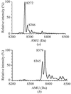
Experimental conditions for the white-beam topography of ribonuclease S crystals were optimized by considering calculated parameters. Images showing some dynamical contrast were thus obtained with minimal radiation damage to the crystals.
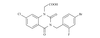
The crystal structure of photoactive yellow protein with a modified chromophore shows the chromophore to be bound in two different conformations. From this crystallographic study and spectroscopic studies, caffeic acid binds the protein in a strained conformation.
PDB reference: caffeic acid PYP, 1kou, r1kousf
A structural and energetic analysis of single and double salt-bridge mutants in barnase reinforces the validity of the double-mutant cycle method used to estimate the strength and cooperativity of molecular interactions in proteins.
Crystal structure of the netropsin–d(CCIICICCII)2 shows a preference of netropsin binding to the target DNA. The conformation differences between the drug-binding and the drug-free portions of the DNA are described.
NDB reference: netropsin–decamer d(CCIICICCII)2 complex, DD0024
Caged ATP was successfully photolyzed at 100–150 K in crystals of thymidylate kinase as assessed by the structural observation of ATP-dependent enzymatic activity. Cryophotolysis of caged compounds, in combination with the use of adequate temperature profiles, opens new possibilities for trapping intermediate states in protein crystals.
X-ray structures of TRAP bound to 53-nucleotide single-stranded RNA containing GAGUU and GAGCC repeats were determined. Comparison of these structures with that of TRAP bound to RNAs containing GAGAU repeats gives insight into the specificity of the TRAP–RNA interaction and suggests that pyrimidine spacer nucleotides bind more strongly owing to the greater increase in entropy upon binding.
The crystal structure of the complex of human recombinant aldose reductase with zenarestat has been solved at 2.5 Å resolution. Zenarestat fits in the hydrophobic active site and induces unique and dramatic conformational changes.
PDB reference: aldose reductase–zenarestat complex, 1iei, r1ieisf

The structure of the RNase-related protein from C. sepium explains its complete lack of ribonuclease activity.
PDB reference: CalsepRRP, 1jy5, r1jy5sf
The rational design and X-ray crystallographic analyses of two symmetrical allosteric effectors of hemoglobin are reported. Both compounds were found to span and link primary and secondary allosteric sites.
PDB reference: TB5-27–Hb, 1k0y, r1k0ysf
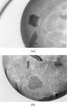
A comparative crystallographic analysis of earth-grown and space-grown crystals of AspRS-1, a large dimeric enzyme having flexible domains, shows that the latter are of enhanced quality. Their mosaicity as well as the electron-density map and the structure model derived from them are improved.
PDB reference: space-grown aspartyl-tRNA synthetase-1, 1l0w, r1l0wsf
The first crystal structure of an endo-β-1,4-glucanase from higher termites refined to 1.4 Å resolution is reported here. The effects of pH changes on the structure of the enzyme are minimal: a structural Ca2+ ion is lost at pH 2.5.

The structure of an endoglucanase from A. niger (EglA) has been determined at 2.1 Å resolution. The mode of inhibition of EglA by palladium chloride was studied by determination of the structure of an EglA–palladium complex.
crystallization papers
The highly soluble domain (1–65) of rat liver P2 has been overexpressed in Escherichia coli as an N-terminal poly-His-tagged protein and crystallized.
Porcine pancreatic elastase (EC 3.4.21.36) was crystallized in complex with a chemically synthesized hybrid squash inhibitor. The optimization of crystallization conditions is described.
Purification, crystallization and preliminary X-ray crystallographic analysis of a C-type lectin-like protein from A. acutus venom.
Experimental strategies for crystallizing a non-covalent abortive complex between the Fpg DNA repair enzyme and DNA containing an abasic site analogue.
A C-terminal nonapeptide truncation improved the morphology and quality of the crystals of isozyme 1 from barley α-amylase for which expression, purification and crystallization is reported
Crystallization and preliminary crystallographic study of cytochrome P450 from B. subtilis, which catalyzes hydroxylation of long-chain fatty acids at the α and β positions using H2O2 as an oxidant, is reported.
Crystals diffracting to 2.45 Å have been obtained of the enzymatically active (3S)-hydroxyacyl-CoA dehydrogenase domain of rat peroxisomal MFE-1.

Crystallization of human S100P in high protein concentration was reported. Crystals diffract to 2.0 Å and belong to space group P41212.
Cytochrome c peroxidase from P. nautica has been crystallized in two different space groups at pH 4.0 and pH 5.3 using sodium citrate and ammonium phosphate as precipitants. Data collection under cryogenic conditions allowed the characterization of the two crystal forms.
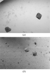
Protein engineering and dynamic light scattering provided the experimental basis to engineer a truncated form of the GTPase Sar1 that behaves well in solution and yields diffraction-quality crystals.
P450 MT2 (CYP121), a novel cytochrome P450 from M. tuberculosis that binds tightly to a range of azole-based antifungal drugs, has been expressed in E. coli, purified and crystallized. The crystals belong to either space group P6122 or P6522. Native crystals diffract to 1.6 Å, while a single-site Hg-derivative crystal diffracts to 2.5 Å resolution.
The selenate reductase from T. selenatis was crystallized by vapour diffusion. Diffraction data from the native and a putative heavy-atom derivative have been recorded.

The successful crystallization and characterization by X-ray diffraction of a recombinant mutant pectate lyase from the hyperthermophilic bacterium T. maritima is described.
The protein biosynthesis elongation factor 2 from yeast has been crystallized and data collected to 2.85 Å. A yeast strain expressing the histidine-tagged factor as the only form of the protein has been constructed.

The C103A mutant of acyl coenzyme A:isopenicillin N, the last enzyme in penicillin G biosynthesis, was purified and crystallized. Crystals of a SeMet derivative were also obtained.
short communications
Analysis of the crystal structure of a trematode haemoglobin displaying very high O2 affinity shows that crystal contacts in two different crystal forms markedly affect the haem-cavity stereochemistry and haem Fe coordination state.
PDB reference: P. epiclitum haemoglobin, 1kfr, r1kfrsf
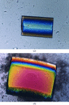
Wild-type cysteine protease precursor from the human pathogen S. pyogenes was obtained in the form of crystalline flakes from which X-ray diffraction data were collected to 3.15 Å resolution at 100 K using synchrotron radiation. The crystals are monoclinic, space group P21, with unit-cell parameters a = 41.6, b = 136.0, c = 156.7 Å, β = 95.7°, and contain four copies of the protein in the asymmetric unit.


