journal menu


issue contents
December 2016 issue
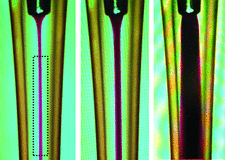
Cover illustration: Improved radiation dose efficiency in solution SAXS using a sheath flow sample environment (Kirby et al., p. 1254). To overcome radiation damage, a major limitation to synchrotron small-angle X-ray scattering analysis, the coflow method has been developed, where the sample flows through the centre of its cell surrounded by a flow of matched buffer. This image shows the stable laminar flow in a coflow cell using a dye solution as a sample and water as a sheath flow, and the effect of increasing the fractional sample flow rate. The dotted box indicates a suggested beam position.
research papers


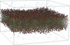
On the interpretation of reflectivity data from lipid bilayers in terms of molecular-dynamics models
A method is presented for producing continuous scattering length density profiles from simulated molecular-dynamics structures of biomembranes for the analysis of reflectometry data from lipid layers.
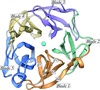
The F-BARx domain of SRGAP2 was produced for crystallization by co-expressing it with the carboxy domains of the protein. The F-BARx crystal structure was determined by a molecular symmetry-constrained systematic search, utilizing the conserved biological symmetry of the F-BAR fold, an approach that is shown to be useful in solving other F-BAR structures.

Open  access
access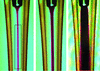
Coflow is a new method for delivering radiation-sensitive biological and other solution-based samples to high-brightness X-ray beamlines that exploits laminar flow to ameliorate radiation-damage limitations and provides a host of practical improvements associated with these types of experiments.

Open access
access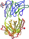
Models of the VL1 glycosylated Fab fragment independently refined from two non-apparent (pseudo) isomorphous crystals show significant differences, allowing the meaning of accuracy in structure description to be revisited, while at the same time inviting reflections about the benefits and boundaries of complex solvent modelling and validation.
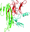
The structure of C. thermocellum arabinofuranosidase 43A reveals how the enzyme acts specifically in the removal of arabinose side chains from arabinoxylan but not from pectic arabinan.
The PERK luminal domain can function as a molecular chaperone to recognize and bind misfolded proteins. The crystal structure of the PERK luminal domain suggests that it may utilize a flexible β-sandwich domain to recognize and interact with a broad range of misfolded proteins.
PDB reference: PERK luminal domain, 5sv7
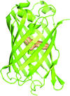
The structures of the fluorescent proteins lanYFP and mNeonGreen from B. lanceolatum are described, providing a structural understanding of the directed evolution process from lanYFP to mNeonGreen. A specific radiation-damage study has been performed for mNeonGreen.
book reviews
Free