
Acta Crystallographica
Section E CRYSTALLOGRAPHIC COMMUNICATIONS


 menu


research communications
| CRYSTALLOGRAPHIC COMMUNICATIONS |
 access
Synthesis, crystal structure and Hirshfeld surface analysis of (E)-5-methoxy-2-[(p-tolylimino)methyl]phenol

aPG and Research Department of Chemistry, Seethalakshmi Ramaswami College, (Affiliated to Bharathidasan University), Tiruchirappalli-620002, Tamil Nadu, India, bDivision of Chemistry, School of Sciences, Faculty of Engineering and Technology, SRM Institute of Science and Technology, Tiruchirappalli-621 105, Tamil Nadu, India, and cDepartment of Chemistry, Indian Institute of Technology, Chennai-620 036, Tamil Nadu, India
*Correspondence e-mail: [email protected]
Single-crystal X-ray diffraction has been used to elucidate the molecular and crystal structures of (E)-5-methoxy-2-[(p-tolylimino)methyl]phenol {systematic name: (E)-5-methoxy-2-[N-(4-methylphenyl)carboximidoyl]phenol}, C15H15NO2. The Schiff base crystallizes in the orthorhombic space group Pca21 and the molecular structure is stabilized by an intramolecular O—H⋯N hydrogen bond, with an O⋯N separation of 2.605 (4) Å and an O—H⋯N angle of 146°. The crystal packing was examined using Hirshfeld surface analysis, which reveals dominant H⋯H contacts make the largest contribution to the intermolecular contacts (51.4%) of the surface, followed by H⋯C/C⋯H (30.9%), H⋯O/O⋯H (12.0%) and H⋯N/N⋯H (2.5%) contacts. The combined crystallographic and Hirshfeld surface analyses provide insight into the molecular organization and intermolecular forces governing the solid-state structure.
Keywords: crystal structure; Schiff base; imine; Hirshfeld surface; hydrogen bond; crystal packing.
CCDC reference: 2480905
1. Chemical context
Schiff bases derived from salicylaldehyde are an important class of compounds owing to their structural diversity and applications in coordination chemistry, molecular recognition and materials chemistry. The azomethine group together with the appropriately positioned phenolic hydroxyl group provides a useful molecular scaffold for structural modification and metal coordination (Govindaraj & Ramanathan, 2014 ). This class of compounds has also attracted attention because of the reported biological potential of Schiff base derivatives, including antimicrobial and antioxidant properties (Govindaraj V et al., 2026c).
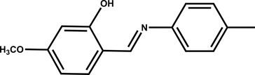
In the present work, (E)-5-methoxy-2-[(p-tolylimino)methyl]phenol was synthesized by condensation of 2-hydroxy-4-methoxybenzaldehyde with p-toluidine. The p-methyl substituent was introduced to provide an electron-donating group while minimizing steric effects near the azomethine linkage. The molecular and crystal structures were determined by single-crystal X-ray diffraction. Hirshfeld surface analysis and energy-framework calculations were subsequently employed to characterize the intermolecular contacts and to assess their contributions to the consolidation of the crystal structure.
2. Structural commentary
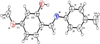
The molecular structure of the title compound, together with the atom-numbering scheme, is shown in Fig. 1. The molecule consists of two benzene rings connected through an azomethine (—CH=N—) linkage. Related Schiff-base structures have been characterized by single-crystal X-ray diffraction (Govindaraj et al., 2026a,b). The C1–C6 ring corresponds to the p-tolyl ring, whereas the C8–C13 ring bears the phenolic hydroxyl and methoxy substituents. The two benzene ring mean planes are inclined at a dihedral angle 49.46 (16)°, indicating a non-planar molecular conformation. The azomethine group adopts a trans configuration, as indicated by the C8—C7—N1—C1 torsion angle of −172.9 (3)°. The torsion angles C2—C1—N1—C7 [−144.2 (3)°] and C6—C1—N1—C7 [41.6 (5)°] further describe the orientation of the p-tolyl ring relative to the imine group. The principal bond lengths are consistent with the expected geometry of the Schiff-base framework. In particular, the N1=C7 bond is 1.284 (4) Å, while the C1—N1 and C7—C8 bonds are 1.420 (4) and 1.432 (5) Å, respectively. The phenolic C13—O2 bond length is 1.345 (4) Å, and the methoxy group is characterized by C11—O1 = 1.358 (4) Å and O-1–C15 = 1.406 (5) Å.
|
Figure 1
The molecular structure of the title compound, showing the atom-numbering scheme and displacement ellipsoids drawn at the 50% probability level. |
3. Supramolecular features
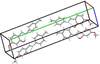
The phenolic hydroxyl group forms an intramolecular O2—H2A⋯N1 hydrogen bond, with an O2⋯N1 distance of 2.605 (4) Å and an O2—H2A⋯N1 angle of 146° (Table 1). This interaction generates a six-membered pseudo-ring and contributes to the stabilization of the molecular conformation. The crystal packing, shown in Fig. 2, is governed mainly by weak intermolecular contacts between neighboring molecules.
|
|||||||||||||||||
|
Figure 2
Crystal packing of the title compound, showing the arrangement of molecules in the crystal viewed along the a axis. |
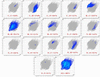
Hirshfeld surface analysis was used to quantify the intermolecular contacts (Dey et al., 2024; Spackman & Jayatilaka, 2009). The Hirshfeld surface has a volume of 311.57 Å3 and a surface area of 299.40 Å3. The surface mappings are shown in Fig. 3. The two-dimensional fingerprint plots (Fig. 4) show that H⋯H contacts make the largest contribution 51.4%), followed by H⋯C/C⋯H contacts (30.9%). The H⋯O/O⋯H contacts contribute 12.0%, while H⋯N/N⋯H contacts contribute 2.5%. Smaller contributions arise from C⋯O/O⋯C (2.4%), C⋯C (0.4%) and N⋯O/O⋯N (0.2%) contacts.

|
Figure 3
Hirshfeld surfaces of the title compound mapped over dnorm, shape-index and curvedness. |
|
Figure 4
Two-dimensional fingerprint plots showing the contributions of the intermolecular contacts in the crystal structure of the title compound. |
4. Database survey
A search of the Cambridge Structural Database (CSD, version 6.01, July 2026 update; Groom et al., 2016) identified the closely related Schiff-base compound (E)-2-[(p-tolylimino)methyl]phenol (refcode CULPOO01; Nieger, 2012). Both compounds exhibit a trans configuration of the azomethine group, with C6—C7—N8—C9 and C8—C7—N1—C1 torsion angles of 179.41 (11) and −172.9 (3)°, respectively. The aromatic rings of the reference compound are nearly coplanar, as indicated by the C7—N8—C9—C10 torsion angle of 172.26 (12)°, whereas the title compound exhibits a more twisted arrangement, with a C2—C1—N1—C7 torsion angle of −144.2 (3)°. Both structures retain an intramolecular O–H⋯N hydrogen bond, with donor–acceptor distances of 2.5963 (14) and 2.605 (4) Å, respectively. The methoxy substituent in the title compound may influence its conformational preferences and crystal packing.
5. Synthesis and crystallization
The title compound was prepared according to the reported procedure of Kudernac et al. (2009) p-toluidine (0.106 g, 1 mmol) and 2-hydroxy-4-methoxybenzaldehyde (0.152 g, 1 mmol) were dissolved in 15 ml of ethanol and the reaction mixture was refluxed for 3 h. After cooling to room temperature, the resulting yellow crystals were collected by filtration and dried under vacuum to give the product in approximately 0.186 g (77%) yield. Single crystals suitable for X-ray diffraction were obtained by allowing the solution to stand at room temperature for approximately 2 days, affording clear light-yellow plate-shaped crystals.
FT–IR (KBr, cm−1): 3546 [ν(O—H)], 1618 [ν(C=N)], 1522 [ν(C=C)], 3021 [ν(C—H)], 2922 [ν(aliphatic C—H, CH3)] and 1288 [ν(C—O)]. 1H NMR (500 MHz, DMSO-d6, δ ppm): 6.54 (d, 1H), 7.28 (d, 1H), 7.25 (d, 1H), 6.56 (d, 1H), 8.85 (s, 1H), 7.51 (d, 1H), 6.48 (d, 1H), 7.23 (s, 1H), 3.80 (s, 3H), 2.32 (s, 3H, Ar–CH3) and 13.81 (s, 1H, OH). HRMS (CH2Cl2): m/z 242.11 [M + H]+·Elemental analysis: calculated for C15H15NO2: C, 74.67; H, 6.27; N, 5.81; O, 13.26%; found: C, 74.66; H, 6.25; N, 5.80; O, 13.26%.
6. Refinement
Details of the crystal data, data collection and refinement are given in Table 2. C-bound H atoms were placed in calculated positions and refined using a riding model, while the O-bound H atom was refined independently.
|
Supporting information
CCDC reference: 2480905
Crystal structure: contains datablock I. DOI: https://doi.org/10.1107/S205698902601056X/tx2117sup1.cif
Structure factors: contains datablock I. DOI: https://doi.org/10.1107/S205698902601056X/tx2117Isup3.hkl
Supporting information file. DOI: https://doi.org/10.1107/S205698902601056X/tx2117Isup3.cml
| C15H15NO2 | Dx = 1.26 Mg m−3 |
| Mr = 241.28 | Mo Kα radiation, λ = 0.71073 Å |
| Orthorhombic, Pca21 | Cell parameters from 6384 reflections |
| a = 7.2146 (7) Å | θ = 2.9–23.7° |
| b = 28.256 (3) Å | µ = 0.08 mm−1 |
| c = 6.2413 (6) Å | T = 300 K |
| V = 1272.3 (2) Å3 | Plate, clear light yellow |
| Z = 4 | 0.34 × 0.22 × 0.08 mm |
| F(000) = 512 |
| Bruker D8 QUEST diffractometer | 3164 independent reflections |
| Radiation source: microfocus sealed tube, INCOATEC IµS 3.0 | 2247 reflections with I > 2σ(I) |
| Multilayer mirror monochromator | Rint = 0.061 |
| Detector resolution: 7.391 pixels mm-1 | θmax = 28.3°, θmin = 2.2° |
| ω and φ scans | h = −9→9 |
| Absorption correction: multi-scan (SADABS; Krause et al., 2015) | k = −36→37 |
| Tmin = 0.972, Tmax = 0.994 | l = −8→8 |
| 29157 measured reflections |
| Refinement on F2 | Hydrogen site location: inferred from neighbouring sites |
| Least-squares matrix: full | H atoms treated by a mixture of independent and constrained refinement |
| R[F2 > 2σ(F2)] = 0.057 | w = 1/[σ2(Fo2) + (0.0623P)2 + 0.323P] where P = (Fo2 + 2Fc2)/3 |
| wR(F2) = 0.151 | (Δ/σ)max = 0.007 |
| S = 1.06 | Δρmax = 0.19 e Å−3 |
| 3164 reflections | Δρmin = −0.17 e Å−3 |
| 167 parameters | Absolute structure: Flack x determined using 786 quotients [(I+)-(I-)]/[(I+)+(I-)] (Parsons et al., 2013) |
| 1 restraint | Absolute structure parameter: 0.2 (6) |
| Primary atom site location: Intrinsic Phasing |
Geometry. All esds (except the esd in the dihedral angle between two l.s. planes) are estimated using the full covariance matrix. The cell esds are taken into account individually in the estimation of esds in distances, angles and torsion angles; correlations between esds in cell parameters are only used when they are defined by crystal symmetry. An approximate (isotropic) treatment of cell esds is used for estimating esds involving l.s. planes. |
Refinement. The diffraction data were processed using SAINT (Bruker, 2016) and absorption correction was applied using SADABS (Krause et al., 2015). The structure was solved by intrinsic phasing using SHELXT 2018/2 and refined by full-matrix least-squares methods against F2 using SHELXL-2019/3 (Sheldrick, 2015a,b). All non-H atoms were refined anisotropically. |
| x | y | z | Uiso*/Ueq | ||
| C1 | 0.5091 (4) | 0.31881 (12) | 0.3712 (6) | 0.0444 (8) | |
| C2 | 0.5694 (5) | 0.35917 (12) | 0.4749 (6) | 0.0518 (9) | |
| H2 | 0.618437 | 0.356993 | 0.612401 | 0.062* | |
| C3 | 0.5574 (5) | 0.40250 (13) | 0.3759 (8) | 0.0585 (10) | |
| H3 | 0.601158 | 0.429228 | 0.446663 | 0.070* | |
| C4 | 0.4811 (5) | 0.40729 (12) | 0.1718 (7) | 0.0558 (10) | |
| C5 | 0.4216 (5) | 0.36685 (12) | 0.0713 (7) | 0.0545 (9) | |
| H5 | 0.372322 | 0.368988 | −0.066013 | 0.065* | |
| C6 | 0.4329 (4) | 0.32304 (13) | 0.1688 (6) | 0.0491 (8) | |
| H6 | 0.389169 | 0.296313 | 0.098084 | 0.059* | |
| C7 | 0.5508 (4) | 0.23666 (12) | 0.3821 (6) | 0.0463 (8) | |
| H7 | 0.590493 | 0.238534 | 0.240619 | 0.056* | |
| C8 | 0.5352 (4) | 0.19109 (11) | 0.4809 (5) | 0.0433 (8) | |
| C9 | 0.5914 (4) | 0.15020 (12) | 0.3729 (6) | 0.0474 (8) | |
| H9 | 0.639037 | 0.153035 | 0.234987 | 0.057* | |
| C10 | 0.5789 (4) | 0.10639 (12) | 0.4623 (6) | 0.0481 (8) | |
| H10 | 0.622562 | 0.080033 | 0.388681 | 0.058* | |
| C11 | 0.5001 (4) | 0.10124 (12) | 0.6659 (6) | 0.0445 (8) | |
| C12 | 0.4384 (5) | 0.14066 (11) | 0.7776 (6) | 0.0462 (8) | |
| H12 | 0.383505 | 0.137234 | 0.911506 | 0.055* | |
| C13 | 0.4595 (4) | 0.18514 (11) | 0.6879 (6) | 0.0441 (8) | |
| C14 | 0.4622 (7) | 0.45488 (14) | 0.0664 (10) | 0.0826 (14) | |
| H14A | 0.536888 | 0.477583 | 0.141530 | 0.124* | |
| H14B | 0.334792 | 0.464653 | 0.070195 | 0.124* | |
| H14C | 0.502843 | 0.452796 | −0.079713 | 0.124* | |
| C15 | 0.4117 (7) | 0.04730 (15) | 0.9405 (7) | 0.0764 (13) | |
| H15A | 0.285977 | 0.058395 | 0.938838 | 0.115* | |
| H15B | 0.479579 | 0.063565 | 1.050459 | 0.115* | |
| H15C | 0.413186 | 0.013907 | 0.968858 | 0.115* | |
| N1 | 0.5117 (4) | 0.27501 (10) | 0.4827 (5) | 0.0481 (7) | |
| O1 | 0.4944 (4) | 0.05613 (9) | 0.7406 (4) | 0.0580 (7) | |
| O2 | 0.4075 (4) | 0.22284 (9) | 0.8050 (4) | 0.0624 (8) | |
| H2A | 0.428 (6) | 0.2502 (14) | 0.727 (5) | 0.094* |
| U11 | U22 | U33 | U12 | U13 | U23 | |
| C1 | 0.0395 (16) | 0.049 (2) | 0.044 (2) | 0.0026 (13) | 0.0062 (17) | 0.0003 (15) |
| C2 | 0.0496 (18) | 0.061 (2) | 0.0447 (19) | −0.0015 (15) | −0.0016 (17) | −0.0056 (19) |
| C3 | 0.0531 (19) | 0.053 (2) | 0.070 (3) | −0.0063 (16) | −0.002 (2) | −0.009 (2) |
| C4 | 0.051 (2) | 0.054 (2) | 0.063 (3) | −0.0020 (16) | 0.004 (2) | 0.0060 (19) |
| C5 | 0.0487 (19) | 0.063 (2) | 0.052 (2) | 0.0035 (16) | 0.0005 (16) | 0.0035 (18) |
| C6 | 0.0488 (17) | 0.055 (2) | 0.044 (2) | −0.0037 (15) | −0.0008 (16) | −0.0053 (15) |
| C7 | 0.0419 (16) | 0.060 (2) | 0.0370 (18) | 0.0005 (14) | −0.0018 (14) | −0.0006 (16) |
| C8 | 0.0412 (16) | 0.0533 (19) | 0.0354 (18) | 0.0018 (13) | −0.0003 (15) | −0.0039 (16) |
| C9 | 0.0445 (17) | 0.060 (2) | 0.0376 (17) | 0.0017 (15) | 0.0043 (15) | −0.0076 (17) |
| C10 | 0.0480 (17) | 0.0534 (19) | 0.0429 (18) | 0.0049 (14) | 0.0013 (16) | −0.0088 (17) |
| C11 | 0.0450 (17) | 0.0473 (19) | 0.0412 (19) | −0.0006 (14) | −0.0057 (16) | −0.0037 (15) |
| C12 | 0.0481 (17) | 0.057 (2) | 0.0332 (17) | 0.0024 (15) | 0.0029 (15) | 0.0009 (15) |
| C13 | 0.0422 (16) | 0.052 (2) | 0.0381 (18) | 0.0042 (14) | 0.0011 (15) | −0.0061 (15) |
| C14 | 0.085 (3) | 0.063 (3) | 0.100 (4) | 0.002 (2) | −0.009 (3) | 0.015 (3) |
| C15 | 0.115 (4) | 0.062 (2) | 0.052 (2) | 0.005 (2) | 0.018 (2) | 0.010 (2) |
| N1 | 0.0457 (14) | 0.0526 (17) | 0.0461 (16) | 0.0012 (12) | 0.0017 (15) | 0.0001 (14) |
| O1 | 0.0721 (18) | 0.0509 (14) | 0.0510 (15) | 0.0016 (12) | 0.0041 (12) | −0.0008 (12) |
| O2 | 0.0848 (19) | 0.0548 (14) | 0.0474 (16) | 0.0107 (14) | 0.0176 (14) | −0.0056 (13) |
| C1—C2 | 1.381 (5) | C9—C10 | 1.361 (5) |
| C1—C6 | 1.383 (5) | C9—H9 | 0.9300 |
| C1—N1 | 1.420 (4) | C10—C11 | 1.399 (5) |
| C2—C3 | 1.374 (5) | C10—H10 | 0.9300 |
| C2—H2 | 0.9300 | C11—O1 | 1.358 (4) |
| C3—C4 | 1.394 (6) | C11—C12 | 1.387 (4) |
| C3—H3 | 0.9300 | C12—C13 | 1.384 (4) |
| C4—C5 | 1.372 (5) | C12—H12 | 0.9300 |
| C4—C14 | 1.503 (5) | C13—O2 | 1.345 (4) |
| C5—C6 | 1.382 (5) | C14—H14A | 0.9600 |
| C5—H5 | 0.9300 | C14—H14B | 0.9600 |
| C6—H6 | 0.9300 | C14—H14C | 0.9600 |
| C7—N1 | 1.284 (4) | C15—O1 | 1.406 (5) |
| C7—C8 | 1.432 (5) | C15—H15A | 0.9600 |
| C7—H7 | 0.9300 | C15—H15B | 0.9600 |
| C8—C9 | 1.398 (4) | C15—H15C | 0.9600 |
| C8—C13 | 1.412 (4) | O2—H2A | 0.92 (5) |
| C2—C1—C6 | 118.8 (3) | C9—C10—C11 | 119.6 (3) |
| C2—C1—N1 | 119.0 (3) | C9—C10—H10 | 120.2 |
| C6—C1—N1 | 121.9 (3) | C11—C10—H10 | 120.2 |
| C3—C2—C1 | 120.3 (4) | O1—C11—C12 | 124.8 (3) |
| C3—C2—H2 | 119.8 | O1—C11—C10 | 114.9 (3) |
| C1—C2—H2 | 119.8 | C12—C11—C10 | 120.2 (3) |
| C2—C3—C4 | 121.5 (4) | C13—C12—C11 | 119.4 (3) |
| C2—C3—H3 | 119.3 | C13—C12—H12 | 120.3 |
| C4—C3—H3 | 119.3 | C11—C12—H12 | 120.3 |
| C5—C4—C3 | 117.4 (3) | O2—C13—C12 | 117.9 (3) |
| C5—C4—C14 | 121.1 (4) | O2—C13—C8 | 120.7 (3) |
| C3—C4—C14 | 121.5 (4) | C12—C13—C8 | 121.4 (3) |
| C4—C5—C6 | 121.8 (4) | C4—C14—H14A | 109.5 |
| C4—C5—H5 | 119.1 | C4—C14—H14B | 109.5 |
| C6—C5—H5 | 119.1 | H14A—C14—H14B | 109.5 |
| C5—C6—C1 | 120.2 (3) | C4—C14—H14C | 109.5 |
| C5—C6—H6 | 119.9 | H14A—C14—H14C | 109.5 |
| C1—C6—H6 | 119.9 | H14B—C14—H14C | 109.5 |
| N1—C7—C8 | 122.1 (3) | O1—C15—H15A | 109.5 |
| N1—C7—H7 | 119.0 | O1—C15—H15B | 109.5 |
| C8—C7—H7 | 119.0 | H15A—C15—H15B | 109.5 |
| C9—C8—C13 | 117.1 (3) | O1—C15—H15C | 109.5 |
| C9—C8—C7 | 120.8 (3) | H15A—C15—H15C | 109.5 |
| C13—C8—C7 | 122.1 (3) | H15B—C15—H15C | 109.5 |
| C10—C9—C8 | 122.3 (3) | C7—N1—C1 | 119.9 (3) |
| C10—C9—H9 | 118.8 | C11—O1—C15 | 119.0 (3) |
| C8—C9—H9 | 118.8 | C13—O2—H2A | 109.5 |
| C6—C1—C2—C3 | −1.4 (5) | C9—C10—C11—O1 | −179.9 (3) |
| N1—C1—C2—C3 | −175.8 (3) | C9—C10—C11—C12 | −1.3 (5) |
| C1—C2—C3—C4 | 1.4 (6) | O1—C11—C12—C13 | 177.1 (3) |
| C2—C3—C4—C5 | −1.3 (6) | C10—C11—C12—C13 | −1.4 (5) |
| C2—C3—C4—C14 | 178.0 (4) | C11—C12—C13—O2 | −176.5 (3) |
| C3—C4—C5—C6 | 1.2 (6) | C11—C12—C13—C8 | 2.8 (5) |
| C14—C4—C5—C6 | −178.0 (4) | C9—C8—C13—O2 | 177.9 (3) |
| C4—C5—C6—C1 | −1.3 (5) | C7—C8—C13—O2 | −3.4 (4) |
| C2—C1—C6—C5 | 1.4 (5) | C9—C8—C13—C12 | −1.3 (4) |
| N1—C1—C6—C5 | 175.6 (3) | C7—C8—C13—C12 | 177.3 (3) |
| N1—C7—C8—C9 | −175.3 (3) | C8—C7—N1—C1 | −172.9 (3) |
| N1—C7—C8—C13 | 6.1 (5) | C2—C1—N1—C7 | −144.2 (3) |
| C13—C8—C9—C10 | −1.5 (4) | C6—C1—N1—C7 | 41.6 (5) |
| C7—C8—C9—C10 | 179.8 (3) | C12—C11—O1—C15 | 3.6 (5) |
| C8—C9—C10—C11 | 2.8 (5) | C10—C11—O1—C15 | −177.8 (3) |
Acknowledgements
The authors thank the Department of Chemistry, Seethalakshmi Ramaswami College, Tiruchirappalli, the Division of Chemistry, School of Sciences, Faculty of Engineering and Technology, SRM Institute of Science and Technology, Tiruchirappalli, and the Department of Chemistry, Indian Institute of Technology Madras, Chennai, for providing the facilities and support required for this work.
References
 Bruker (2016). SAINT. Bruker AXS Inc., Madison, Wisconsin, USA.
Google Scholar
Dey, P., Islam, S., Das, P. & Seth, S. K. (2024). J. Mol. Struct. 1296, 136820.
CrossRef
Google Scholar
Govindaraj, V. & Ramanathan, S. (2014). Turk. J. Chem. 38, 521–530.
CrossRef
CAS
Google Scholar
Govindaraj, V., Mathiazhagan, P., Ramasamy, S., Packiam, E. J., Ramanathan, S., Dhanabal, D. & Chandrasekran, K. (2026a). Chem. Pap. 80, 7973–7987.
CrossRef
CAS
Google Scholar
Govindaraj, V., Pradeepalakshmi, G. M., Packiam, E. J. & Ramanathan, S. (2026b). Vietnam J. Chem. e70155.
Google Scholar
Govindaraj, V., Pradeepalakshmi, G. M., Ramasamy, S. & Ramanathan, S. (2026c). Bull. Chem. Soc. Ethiop. 40, 2639–2653.
CrossRef
Google Scholar
Groom, C. R., Bruno, I. J., Lightfoot, M. P. & Ward, S. C. (2016). Acta Cryst. B72, 171–179.
Web of Science
CrossRef
IUCr Journals
Google Scholar
Hübschle, C. B., Sheldrick, G. M. & Dittrich, B. (2011). J. Appl. Cryst. 44, 1281–1284.
Web of Science
CrossRef
IUCr Journals
Google Scholar
Krause, L., Herbst-Irmer, R., Sheldrick, G. M. & Stalke, D. (2015). J. Appl. Cryst. 48, 3–10.
Web of Science
CSD
CrossRef
ICSD
CAS
IUCr Journals
Google Scholar
Kudernac, T., Sändig, N., Fernández Landaluce, T., van Wees, B. J., Rudolf, P., Katsonis, N., Zerbetto, F. & Feringa, B. L. (2009). J. Am. Chem. Soc. 131, 15655–15659.
CrossRef
CAS
Google Scholar
Neiger, M. (2012). CSD Communication (refcode CULPOO01). CCDC, Cambridge, England.
Google Scholar
Parsons, S., Flack, H. D. & Wagner, T. (2013). Acta Cryst. B69, 249–259.
Web of Science
CSD
CrossRef
CAS
IUCr Journals
Google Scholar
Sheldrick, G. M. (2015a). Acta Cryst. A71, 3–8.
Web of Science
CrossRef
IUCr Journals
Google Scholar
Sheldrick, G. M. (2015b). Acta Cryst. C71, 3–8.
Web of Science
CrossRef
IUCr Journals
Google Scholar
Spackman, M. A. & Jayatilaka, D. (2009). CrystEngComm 11, 19–32.
Web of Science
CrossRef
CAS
Google Scholar
This is an open-access article distributed under the terms of the Creative Commons Attribution (CC-BY) Licence, which permits unrestricted use, distribution, and reproduction in any medium, provided the original authors and source are cited.
| CRYSTALLOGRAPHIC COMMUNICATIONS |
access