
Acta Crystallographica
Section E CRYSTALLOGRAPHIC COMMUNICATIONS


 menu


early career research
| CRYSTALLOGRAPHIC COMMUNICATIONS |
 access
Crystal structure of (R)-3-fluoropyrrolidine hydrochloride

aTulane University, Department of Chemistry, 6400 Freret Street, 2015 Percival Stern Hall, New Orleans, LA, 70118, USA, and bDepartment of Chemical & Biological Engineering, Northwestern University, Evanston, IL, 60208, USA
*Correspondence e-mail: [email protected]
This article is part of the collection Early Career Scientists in Structural Science.
The title compound [also called (R)-3-fluoropyrrolidin-1-ium chloride], C4H9FN+·Cl−, crystallizes in the monoclinic Sohncke space group P21 (#4). The asymmetric unit contains one 3-fluoropyrrolidinium cation and one chloride anion. The pyrrolidinium ring adopts a puckered conformation, and the fluorine substituent is bonded to C3 at a distance of 1.396 (3) Å. In the crystal, the ammonium N—H groups form hydrogen bonds with the chloride anions, creating an extended hydrogen-bonded arrangement. Although 3-fluoropyrrolidinium derivatives have been investigated as chiral organic components in hybrid materials, including magnetic and ferroelectric systems, the crystallographic characterization of the hydrochloride salt itself has not been reported.
Keywords: crystal structure; (R)-3-fluoropyrrolidine hydrochloride; hydrochloride salt; (R)-3-fluoropyrrolidinium chloride.
CCDC reference: 2598580
1. Chemical context
In recent years, chiral 3-fluoropyrrolidinium cations have attracted interest as the organic components in organic–inorganic hybrid materials with magnetic and ferroelectric properties. The addition of (R)- and (S)-3-fluoropyrrolidinium in cyanido-bridged dimetallic materials has led to the creation of homochiral multiferroic substances that exhibit ferroelectric and ferroelastic behavior (Mao et al., 2022 ).
(R)- and (S)-3-fluoropyrrolidinium cations have also been integrated into quasi-two-dimensional copper(II) chloride hybrid perovskites, materials, which, with the molecule's addition, exhibit magnetic chirality (Zhang et al., 2026). In addition, the cation has been used in the context of alkaline-earth-metal hybrid perovskites, one of which being [(R)-3-fluoropyrrolidinium]Ba(ClO4)3 where molecular fluorination demonstrated an enhancement of several ferroelectric properties relative to the corresponding unsubstituted pyrrolidinium material (Zhou et al., 2025). (R)-3-Fluoropyrrolidinium has also been used to create hexagonal perovskite-type manganese(II) chloride ferroelectrics (Ai et al., 2019).
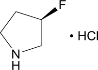
Despite the incorporation of 3-fluoropyrrolidinium in many chiral organic–inorganic hybrid materials, the hydrochloride salt of (R)-3-fluoropyrrolidine [also known as (R)-3-fluoropyrrolidinium chloride], does not have a publicly reported crystal structure. The structure presented here therefore provides a crystallographic characterization of the organic salt, which enables the conformation, stereochemistry, and intermolecular interactions of the 3-fluoropyrrolidinium cation to be independently examined, not in the context of an inorganic hybrid framework.
2. Structural commentary
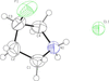
The title compound crystallizes in the monoclinic Sohncke space group P21 (#4). The asymmetric unit contains one (R)-3-fluoropyrrolidinium cation and one chloride anion (Fig. 1). Two formula units compose the unit cell. The pyrrolidinium ring has a non-planarenvelope conformation, with C3 out of plane. The C1—C2—C3—C4 and C2—C3—C4—N1 torsion angles are 36.2 (2)° and −33.8 (2)°, respectively, indicating that the five-membered ring is puckered rather than planar. The C3 atom is bonded to fluorine, with a bond length of 1.396 (3) Å, and the geometry around this carbon is approximately tetrahedral with F1—C3—C2, F1—C3—C4, and C2—C3—C4 angles of 109.2 (2), 109.1 (2), and 103.98 (16)°, respectively. The protonated nitrogen also has approximately tetrahedral geometry.
|
Figure 1
Molecular structure of the title compound with displacement ellipsoids drawn at the 50% probability level. |
3. Supramolecular features
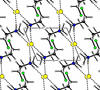
The cations and chloride anions are connected through N—H⋯Cl hydrogen bonds (Fig. 2, Table 1). Each pyrrolidinium cation contains two N—H groups, which both participate in hydrogen bonding with the chloride anions. Nearly linear, these interactions connect neighboring ions and allow for an extended hydrogen-bonded structure throughout the crystal.
|
||||||||||||||||||||||
 .
|
Figure 2
Hydrogen-bonding network in the title compound viewed down the b axis, with Cl− ions shown in yellow. |
4. Database survey
Unfluorinated pyrrolidine hydrochloride crystallizes in the orthorhombic space group Pnma (#62) with V/Z = 155.877 Å3 (Giglmeier et al., 2009). Fluorination causes V/Z to increase to 159.532 Å3 in (R)-3-fluoropyrrolidine hydrochloride, though thermal expansion may account for some of the difference in volume as the crystal structure of pyrrolidine hydrochloride was collected at 200 K whereas the structure of (R)-3-fluoropyrrolidine hydrochloride was collected at 295 K.
5. Synthesis and crystallization
1 mmol NiCl2·6H2O (Thermo Scientific) and 2 mmol (R)-3-fluoropyrrolidine hydrochloride (Ambeed) were dissolved in 0.25 mL of methanol. Crystals were obtained by slow vapor diffusion of diethyl ether into the solution. After five days, the crystals were rinsed with the antisolvent several times and dried under vacuum. The obtained crystals were found to be recrystallized (R)-3-fluoropyrrolidine hydrochloride; an organic-inorganic hybrid material containing both nickel chloride and (R)-3-fluoropyrrolidinium did not form.
6. Refinement
Crystal data, data collection and structure refinement details are summarized in Table 2. Carbon-bonded hydrogen atoms were refined using a riding model with the C—H bond length allowed to vary using the HFIX 24 (two sp3 hydrogens for C1, C2, and C4) or HFIX 14 (one sp3 hydrogen for C3) commands in SHELXL. The positions of the nitrogen-bonded hydrogen atoms (H1A and H1B) were allowed to freely refine, and their isotropic thermal parameters were set to 1.2Ueq(N). The structure was refined using the OLEX2 GUI (Dolomanov et al., 2009).
|
Supporting information
CCDC reference: 2598580
Crystal structure: contains datablock I. DOI: https://doi.org/10.1107/S2056989026011199/jp2036sup1.cif
Structure factors: contains datablock I. DOI: https://doi.org/10.1107/S2056989026011199/jp2036Isup2.hkl
Supporting information file. DOI: https://doi.org/10.1107/S2056989026011199/jp2036Isup3.smi
Supporting information file. DOI: https://doi.org/10.1107/S2056989026011199/jp2036Isup4.cml
| C4H9FN+·Cl− | F(000) = 132 |
| Mr = 125.57 | Dx = 1.307 Mg m−3 |
| Monoclinic, P21 | Mo Kα radiation, λ = 0.71073 Å |
| a = 5.1390 (1) Å | Cell parameters from 9897 reflections |
| b = 10.6704 (2) Å | θ = 3.5–28.2° |
| c = 6.3479 (1) Å | µ = 0.50 mm−1 |
| β = 113.564 (1)° | T = 295 K |
| V = 319.06 (1) Å3 | Prism, brown |
| Z = 2 | 0.38 × 0.21 × 0.16 mm |
| Bruker D8 Quest diffractometer | 1857 independent reflections |
| Radiation source: sealed tube | 1781 reflections with I > 2σ(I) |
| Flat graphite monochromator | Rint = 0.024 |
| Detector resolution: 7.40 pixels mm-1 | θmax = 30.1°, θmin = 3.5° |
| ω and phi scans | h = −7→7 |
| Absorption correction: multi-scan (SADABS; Krause et al., 2015) | k = −15→15 |
| Tmin = 0.932, Tmax = 1.000 | l = −8→8 |
| 18053 measured reflections |
| Refinement on F2 | Hydrogen site location: mixed |
| Least-squares matrix: full | H atoms treated by a mixture of independent and constrained refinement |
| R[F2 > 2σ(F2)] = 0.025 | w = 1/[σ2(Fo2) + (0.040P)2 + 0.016P] where P = (Fo2 + 2Fc2)/3 |
| wR(F2) = 0.068 | (Δ/σ)max < 0.001 |
| S = 1.07 | Δρmax = 0.23 e Å−3 |
| 1857 reflections | Δρmin = −0.13 e Å−3 |
| 74 parameters | Absolute structure: Flack x determined using 806 quotients [(I+)-(I-)]/[(I+)+(I-)] (Parsons et al. (2013) |
| 1 restraint | Absolute structure parameter: −0.017 (16) |
| Primary atom site location: dual |
Geometry. All esds (except the esd in the dihedral angle between two l.s. planes) are estimated using the full covariance matrix. The cell esds are taken into account individually in the estimation of esds in distances, angles and torsion angles; correlations between esds in cell parameters are only used when they are defined by crystal symmetry. An approximate (isotropic) treatment of cell esds is used for estimating esds involving l.s. planes. |
| x | y | z | Uiso*/Ueq | ||
| Cl1 | 0.50723 (8) | 0.66790 (4) | 0.02568 (7) | 0.05125 (14) | |
| F1 | 0.6093 (3) | 0.4266 (3) | 0.5424 (3) | 0.1009 (6) | |
| N1 | 0.0917 (3) | 0.54125 (16) | 0.1987 (3) | 0.0477 (3) | |
| H1A | 0.213 (6) | 0.577 (2) | 0.153 (4) | 0.057* | |
| H1B | −0.073 (6) | 0.574 (2) | 0.140 (4) | 0.057* | |
| C1 | 0.0736 (5) | 0.4059 (3) | 0.1332 (4) | 0.0642 (5) | |
| H1C | −0.120 (5) | 0.3842 (6) | 0.027 (3) | 0.077* | |
| H1D | 0.204 (3) | 0.3871 (5) | 0.0589 (18) | 0.077* | |
| C2 | 0.1553 (6) | 0.3349 (2) | 0.3517 (5) | 0.0704 (6) | |
| H2A | 0.259 (3) | 0.264 (2) | 0.3500 (5) | 0.084* | |
| H2B | −0.005 (5) | 0.3108 (8) | 0.3758 (9) | 0.084* | |
| C3 | 0.3344 (4) | 0.4251 (3) | 0.5356 (4) | 0.0625 (5) | |
| H3 | 0.3345 (4) | 0.4034 (7) | 0.686 (5) | 0.075* | |
| C4 | 0.1987 (5) | 0.5491 (2) | 0.4540 (4) | 0.0609 (5) | |
| H4A | 0.335 (3) | 0.6162 (15) | 0.5129 (14) | 0.073* | |
| H4B | 0.044 (3) | 0.5631 (4) | 0.5017 (11) | 0.073* |
| U11 | U22 | U33 | U12 | U13 | U23 | |
| Cl1 | 0.04294 (19) | 0.0534 (2) | 0.0592 (2) | 0.00460 (17) | 0.02232 (15) | 0.0093 (3) |
| F1 | 0.0422 (6) | 0.1661 (18) | 0.0836 (11) | 0.0214 (10) | 0.0139 (7) | 0.0195 (13) |
| N1 | 0.0365 (6) | 0.0502 (7) | 0.0559 (8) | 0.0036 (6) | 0.0179 (6) | 0.0085 (6) |
| C1 | 0.0690 (12) | 0.0578 (10) | 0.0551 (11) | −0.0026 (10) | 0.0135 (9) | −0.0105 (10) |
| C2 | 0.0733 (15) | 0.0504 (11) | 0.0848 (17) | 0.0087 (10) | 0.0289 (13) | 0.0116 (10) |
| C3 | 0.0512 (10) | 0.0869 (15) | 0.0486 (10) | 0.0086 (11) | 0.0189 (9) | 0.0120 (10) |
| C4 | 0.0596 (11) | 0.0633 (12) | 0.0561 (11) | −0.0061 (9) | 0.0193 (9) | −0.0136 (9) |
| F1—C3 | 1.396 (3) | C2—H2A | 0.93 (3) |
| N1—H1A | 0.88 (3) | C2—H2B | 0.93 (3) |
| N1—H1B | 0.85 (3) | C2—C3 | 1.509 (4) |
| N1—C1 | 1.496 (3) | C3—H3 | 0.98 (3) |
| N1—C4 | 1.491 (3) | C3—C4 | 1.489 (4) |
| C1—H1C | 0.98 (2) | C4—H4A | 0.97 (2) |
| C1—H1D | 0.98 (2) | C4—H4B | 0.97 (2) |
| C1—C2 | 1.486 (4) | ||
| H1A—N1—H1B | 113 (2) | H2A—C2—H2B | 108.9 |
| C1—N1—H1A | 107.8 (16) | C3—C2—H2A | 110.8 |
| C1—N1—H1B | 109.6 (17) | C3—C2—H2B | 110.8 |
| C4—N1—H1A | 109.4 (15) | F1—C3—C2 | 109.2 (2) |
| C4—N1—H1B | 108.6 (18) | F1—C3—H3 | 111.4 |
| C4—N1—C1 | 108.14 (16) | F1—C3—C4 | 109.1 (2) |
| N1—C1—H1C | 110.6 | C2—C3—H3 | 111.4 |
| N1—C1—H1D | 110.6 | C4—C3—C2 | 103.98 (16) |
| H1C—C1—H1D | 108.7 | C4—C3—H3 | 111.4 |
| C2—C1—N1 | 105.65 (18) | N1—C4—H4A | 110.9 |
| C2—C1—H1C | 110.6 | N1—C4—H4B | 110.9 |
| C2—C1—H1D | 110.6 | C3—C4—N1 | 104.23 (18) |
| C1—C2—H2A | 110.8 | C3—C4—H4A | 110.9 |
| C1—C2—H2B | 110.8 | C3—C4—H4B | 110.9 |
| C1—C2—C3 | 104.7 (2) | H4A—C4—H4B | 108.9 |
| F1—C3—C4—N1 | 82.6 (2) | C1—C2—C3—C4 | 36.2 (2) |
| N1—C1—C2—C3 | −24.0 (2) | C2—C3—C4—N1 | −33.8 (2) |
| C1—N1—C4—C3 | 19.2 (2) | C4—N1—C1—C2 | 3.1 (2) |
| C1—C2—C3—F1 | −80.2 (2) |
| D—H···A | D—H | H···A | D···A | D—H···A |
| N1—H1A···Cl1 | 0.87 (3) | 2.20 (3) | 3.0770 (18) | 178 (2) |
| N1—H1B···Cl1i | 0.85 (3) | 2.22 (3) | 3.0686 (18) | 174 (2) |
| Symmetry code: (i) x−1, y, z. |
Acknowledgements
Data were collected in the Joel T. Mague X-ray Crystallography Laboratory at the Department of Chemistry, Tulane University.
Funding information
Funding for this research was provided by: Louisiana Board of Regents (contract No. LEQSF(2024-27)-RD-A-28).
References
 Ai, Y., Chen, X.-G., Shi, P.-P., Tang, Y.-Y., Li, P.-F., Liao, W.-Q. & Xiong, R.-G. (2019). J. Am. Chem. Soc. 141, 4474–4479.
Web of Science
CSD
CrossRef
CAS
PubMed
Google Scholar
Bruker (2016). SAINT. Bruker AXS Inc., Madison, Wisconsin, USA.
Google Scholar
Bruker (2026). BIS. Bruker AXS Inc., Madison, Wisconsin, USA.
Google Scholar
Dolomanov, O. V., Bourhis, L. J., Gildea, R. J., Howard, J. A. K. & Puschmann, H. (2009). J. Appl. Cryst. 42, 339–341.
Web of Science
CrossRef
CAS
IUCr Journals
Google Scholar
Giglmeier, H., Kerscher, T., Klüfers, P. & Mayer, P. (2009). Acta Cryst. E65, o592.
Web of Science
CSD
CrossRef
IUCr Journals
Google Scholar
Krause, L., Herbst-Irmer, R., Sheldrick, G. M. & Stalke, D. (2015). J. Appl. Cryst. 48, 3–10.
Web of Science
CSD
CrossRef
ICSD
CAS
IUCr Journals
Google Scholar
Mao, Y., Chen, X.-G., Gu, Z., Zhang, Z.-X., Song, X.-J., Gu, N. & Xiong, R. G. (2022). Angew. Chem. Int. Ed. 61, e202204135.
CSD
CrossRef
Google Scholar
Parsons, S., Flack, H. D. & Wagner, T. (2013). Acta Cryst. B69, 249–259.
Web of Science
CSD
CrossRef
CAS
IUCr Journals
Google Scholar
Sheldrick, G. M. (2015a). Acta Cryst. A71, 3–8.
Web of Science
CrossRef
IUCr Journals
Google Scholar
Sheldrick, G. M. (2015b). Acta Cryst. C71, 3–8.
Web of Science
CrossRef
IUCr Journals
Google Scholar
Spek, A. L. (2020). Acta Cryst. E76, 1–11.
Web of Science
CrossRef
IUCr Journals
Google Scholar
Westrip, S. P. (2010). J. Appl. Cryst. 43, 920–925.
Web of Science
CrossRef
CAS
IUCr Journals
Google Scholar
Zhang, Z., Xu, M., Gonzalez Jimenez, J. L., Zhang, S., Xie, W., Xu, X. & Straus, D. B. (2026). JACS 148, 33895–33904.
CSD
CrossRef
CAS
Google Scholar
Zhou, Q.-F., Pan, L., Ni, H.-F., Ye, L.-K., Zhao, Y.-P., Luo, Q.-K., Teri, G., Luo, J.-Q., Fu, D.-W., Zhang, Z.-X., Shi, C. & Zhang, Y. (2025). Angew. Chem. Int. Ed. 64, e202509006.
CrossRef
Google Scholar
This is an open-access article distributed under the terms of the Creative Commons Attribution (CC-BY) Licence, which permits unrestricted use, distribution, and reproduction in any medium, provided the original authors and source are cited.
| CRYSTALLOGRAPHIC COMMUNICATIONS |