
Acta Crystallographica Section F
Acta Crystallographica
Section F STRUCTURAL BIOLOGY COMMUNICATIONS
Acta Crystallographica
Section F STRUCTURAL BIOLOGY COMMUNICATIONS


 menu
|
|
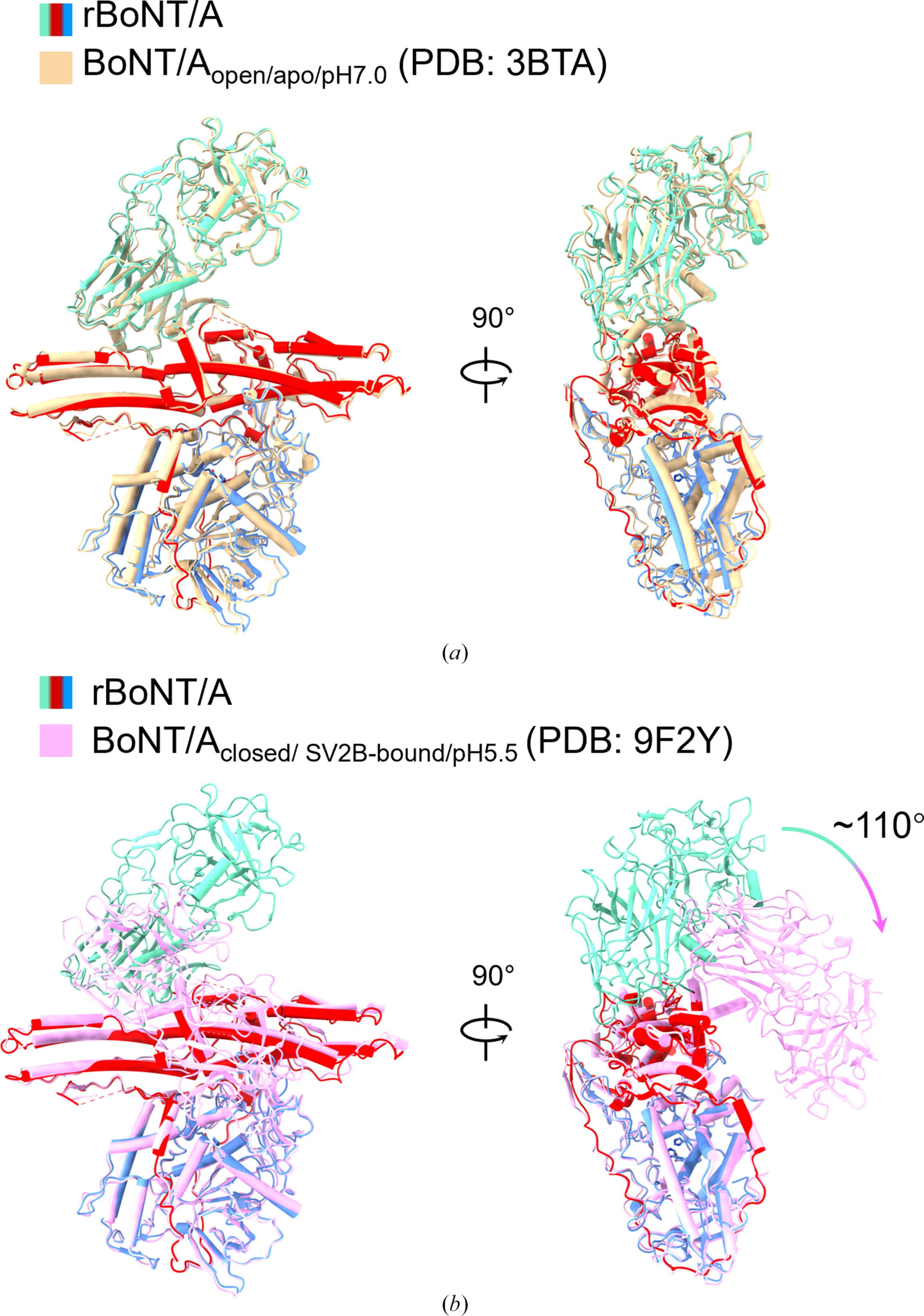
Figure 2
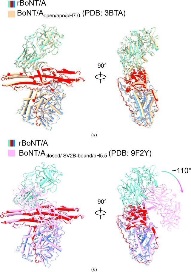
Structural comparison of rBoNT/A with BoNT/A in its open and closed conformations. (a) Side view of the superimposition of rBoNT/A (LC in blue, HN in red and HC in green) and BoNT/Aopen/apo/pH7.0 (PDB entry 3bta, wheat) when they are aligned at the LC domain. (b) Side view of the superimposition of rBoNT/A (LC in blue, HN in red and HC in green) and BoNT/Aclosed/SV2B-bound/pH5.5 (PDB entry 9f2y, pink) when they are aligned at the LC domain. Green–pink arrows indicate the rotation of the HC domain. |
| STRUCTURAL BIOLOGY COMMUNICATIONS |
ISSN: 2053-230X
Open  access