Acta Crystallographica Section F
Acta Crystallographica
Section F STRUCTURAL BIOLOGY COMMUNICATIONS
Acta Crystallographica
Section F STRUCTURAL BIOLOGY COMMUNICATIONS


 menu
|
|
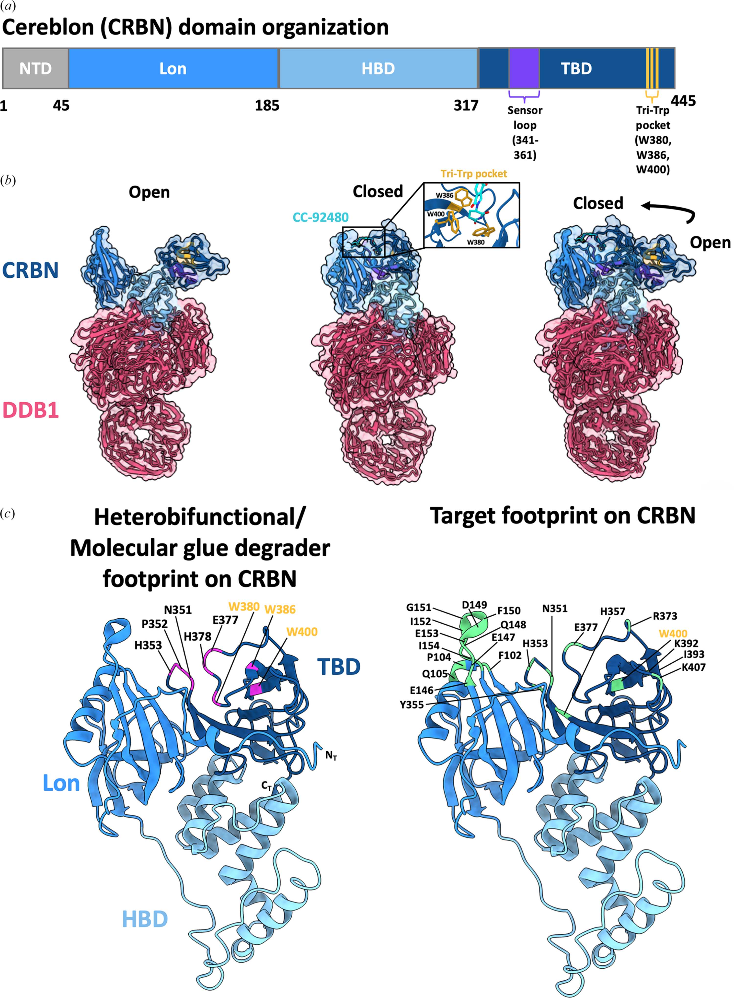
Figure 1
Molecular architecture of cereblon (CRBN) including degrader- and target-binding epitopes. (a) Domain organization of CRBN. Lon/NTD, N-terminal domain; HBD, helical bundle domain; TBD, thalidomide-binding domain. (b) Surface models from the cryo-EM structures of apo/open (PDB entry 8cvp) and degrader-bound/closed (CC-92480; PDB entry 8d7u) states. The transition from the open to closed form in CRBN–DDB1 is shown. Insets show the tri-Trp pocket (Trp380, Trp 386 and Trp400) in orange and sensor-loop (341–361) residues in purple. (c) Heterobifunctional and molecular-glue degrader and target-interacting residues on CRBN from the case studies discussed in this review. Interacting residues highlighted on the CRBN Lon and thalidomide-binding domains from (b) are based on PDB-deposited cryo-EM/X-ray crystallographic ternary-complex structures. NT and CT denote the N-terminus and C-terminus, respectively. Tri-tryptophan residues are highlighted in yellow. |
| STRUCTURAL BIOLOGY COMMUNICATIONS |
ISSN: 2053-230X
 access