

 menu
|
|
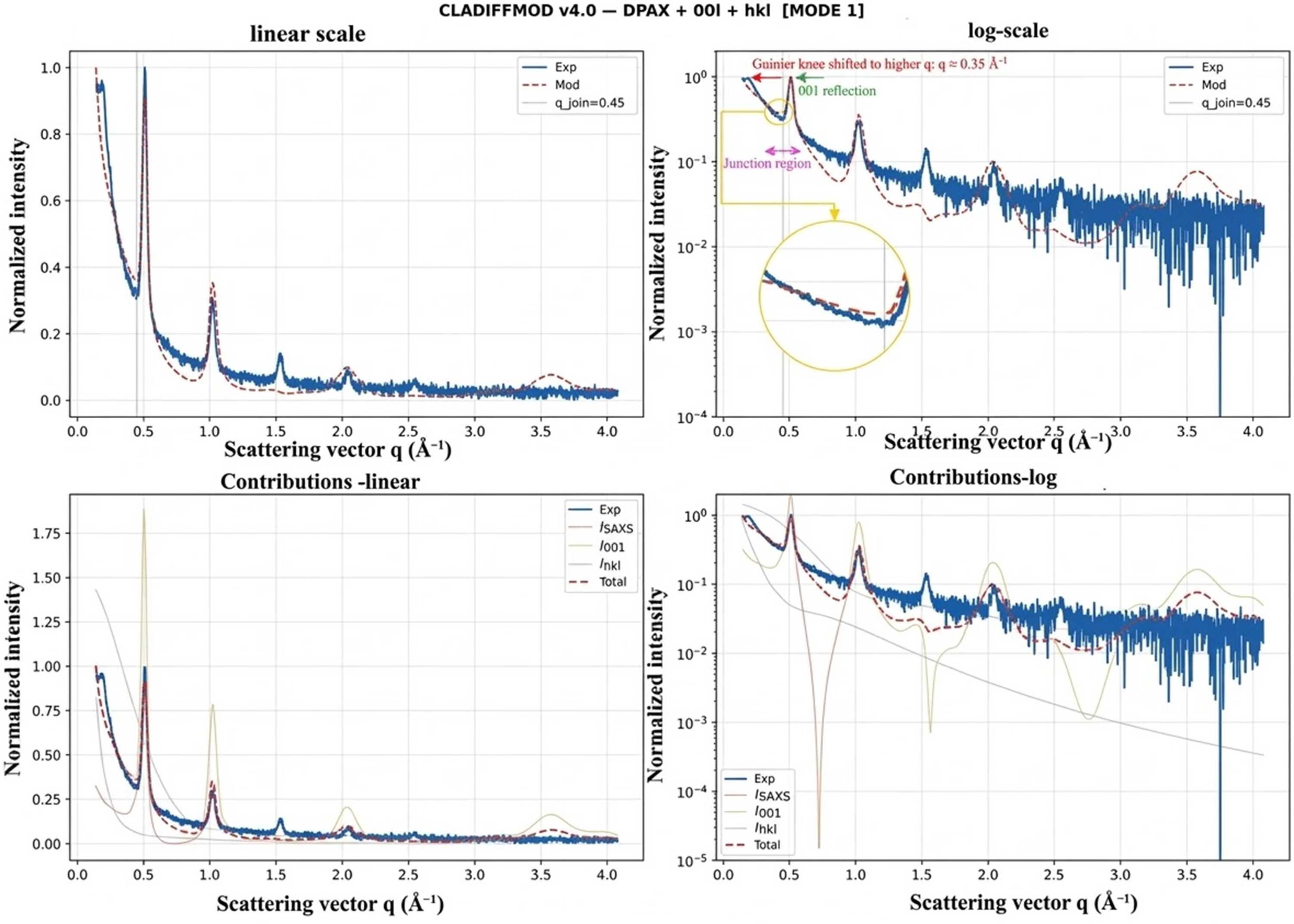
Figure 3
Comparison of experimental and calculated scattering intensities for samples S1 and S4 (43% RH, identical hydration state, different particle sizes). While the basal reflections remain unchanged, significant differences are observed in the low-q region. The model reproduces these variations through changes in the platelet size distribution, confirming that morphological parameters primarily control the SAXS regime. |
 | JOURNAL OF APPLIED CRYSTALLOGRAPHY |
ISSN: 1600-5767
 access