

 menu
|
|
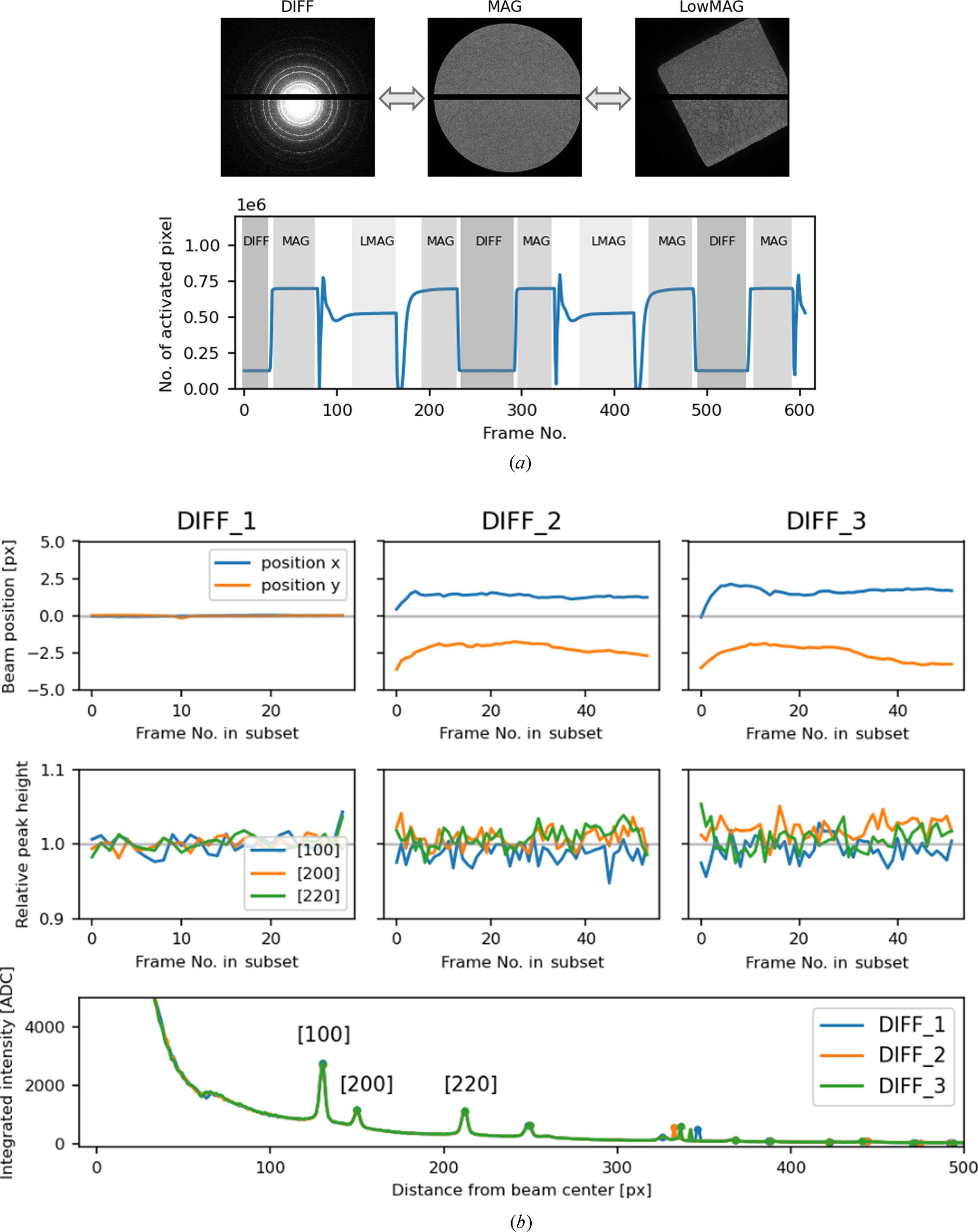
Figure 4
Beam focus reproducibility during magnification-mode switching. (a) (Top) Representative images acquired in DIFF, MAG and LowMAG modes, which are typically used for diffraction measurements, fine crystal alignment and crystal searching, respectively. (Bottom) Number of pixels detecting more than two electrons per frame in a continuous TEM image series, during which TEM modes were switched intermittently. As the illumination area depends on the TEM mode, the active mode can be identified on a per-frame basis, excluding intermediate transition frames. (b) Temporal evolution of the direct-beam position (top) and diffraction peak intensity (middle) for three frame subsets acquired in DIFF mode. The three major peaks in the azimuth integration profile (100, 200 and 220; bottom) are shown for comparison. These profiles mostly overlap, indicating stable focus reproduction despite multiple lens-current changes during mode switching. |
 | JOURNAL OF APPLIED CRYSTALLOGRAPHY |
ISSN: 1600-5767
 access