


 menu
|
|
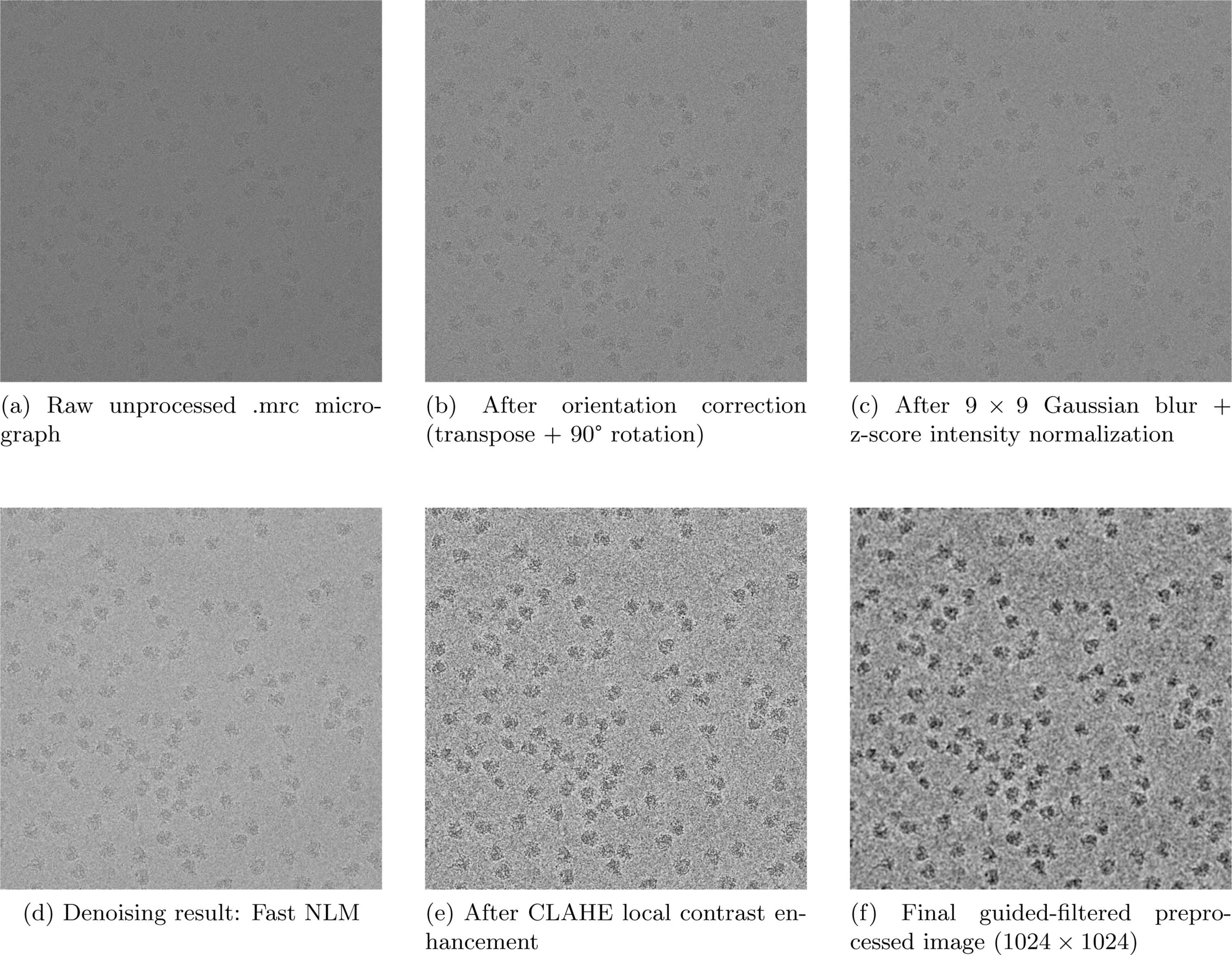
Figure 2
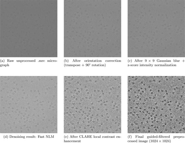
(a) Original raw cryo-EM micrograph loaded from .mrc file. (b) Micrograph after transposition and 90° rotation for orientation rectification. (c) Normalized image after Gaussian smoothing and z-score intensity scaling to 0–255. (d) Denoised output combining fast non-local means (NLM) and Wiener filtering with 9 × 9 Gaussian kernel. (e) Contrast-adjusted image processed by CLAHE. (f) Final preprocessed image after guided filtering (Wiener result as guidance map), resized to 1024 × 1024 for model input. |
IUCrJ
ISSN: 2052-2525
CRYO | EM
Open  access