


 menu
|
|
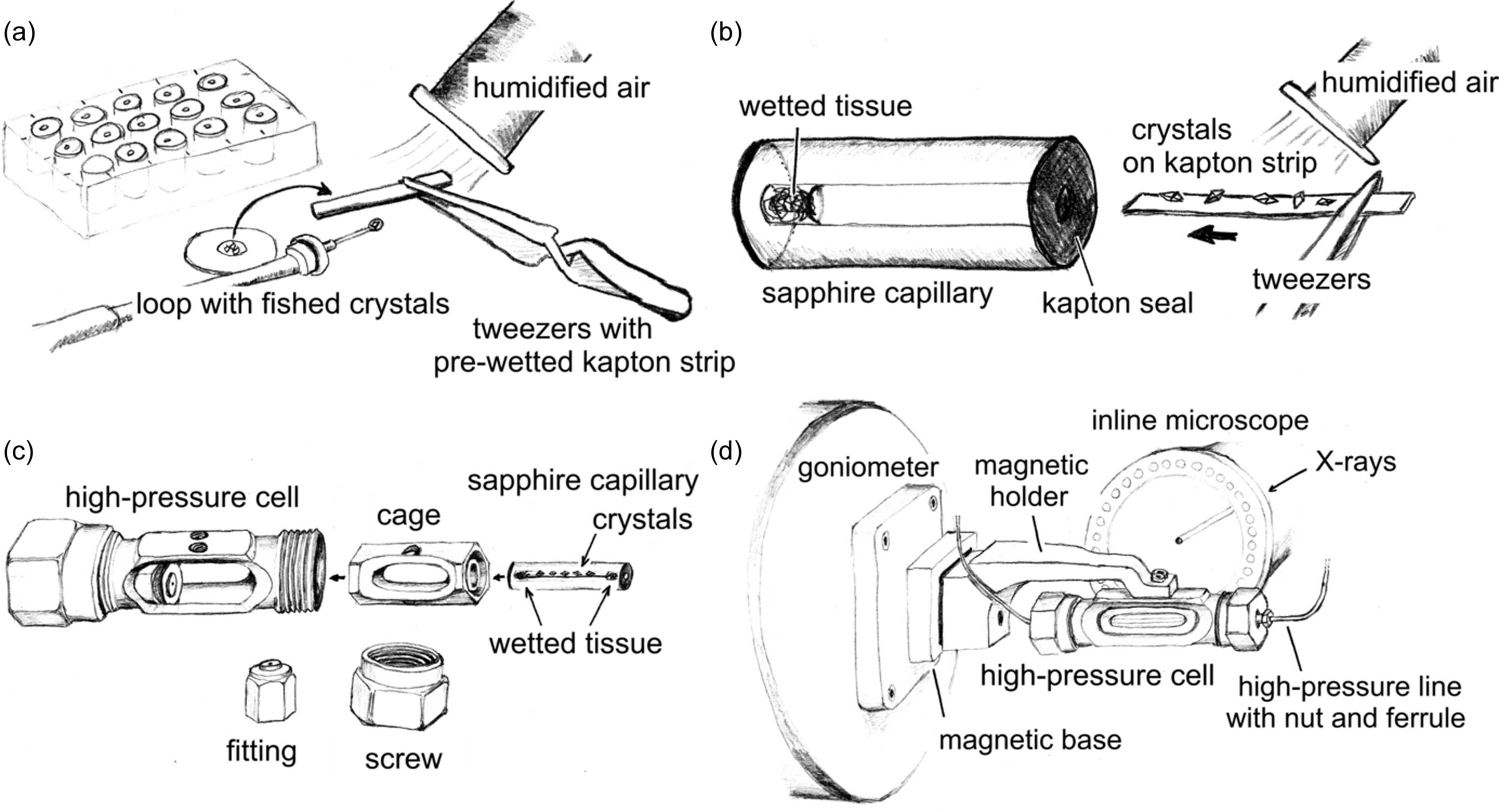
Figure 1
General procedure of an HP experiment with the cell. (a) Multiple protein crystals are transferred onto a pre-wetted Kapton support strip (1100 × 650 × 75 µm) in a stream of humidified air to prevent crystal dehydration. The application of protective oil (e.g. NVH oil) is recommended at this stage for dehydration-sensitive crystals. (b) The Kapton strip is inserted into the sapphire capillary, which already contains a small piece of wet paper tissue serving as a liquid reservoir at the opposite end. Following the Kapton strip, a second piece of paper tissue is inserted into the capillary from the same side, which is then immediately soaked with reservoir solution. (c) Subsequently, the capillary with the crystals is inserted into the central steel cage of the HP cell. The capillary with the Kapton strip can now be manually rotated to orient the crystals favorably with respect to the opening window of the cell and point towards the inline sample-viewing microscope when mounted later on the goniometer. Now, the cell assembly is completed and the screws are tightened. (d) The HP cell is attached to the goniometer of the beamline using a magnetic holder and the crystals are optically centered in the 26 keV X-ray beam. Prior to data collection, the cell is pressurized via nickel-clad fused-silica lines on both sides, which are connected to a double-stage air-driven helium compressor. |
IUCrJ
ISSN: 2052-2525
BIOLOGY | MEDICINE
Open  access