


 menu
|
|
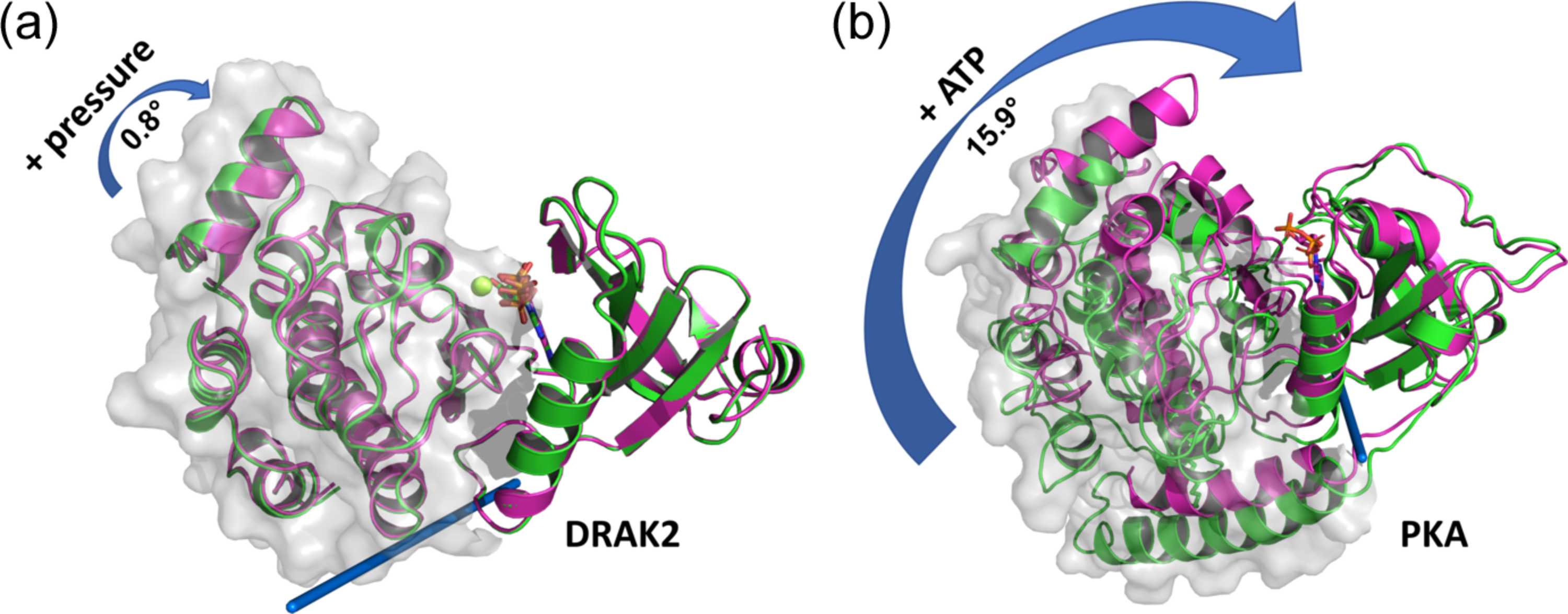
Figure 5
Domain movements in kinases. Comparison of (a) the pressure-induced domain movement of DRAK2 (by 0.8°) and (b) the domain movement observed upon ATP binding to the mammalian cAMP-dependent protein kinase A (PKA) (by 15.9°). Central cleft closure is observed upon ATP binding to PKA and – to a much smaller extent – upon pressure perturbation in DRAK2. The structures at atmospheric pressure and the corresponding nucleotide-free form are shown in green, whereas the 110 MPa structure and the ATP-bound state are colored pink. The structures are superimposed on their N-lobes (right-hand domains) and the C-lobes of the reference states are highlighted as light-gray surfaces. The rotation axes of the hinge movements as determined via the DynDom server (Girdlestone & Hayward, 2016  ) are represented as blue rods. PKA structures 1cmk (free form) and 1atp (ATP-bound form) (Zheng et al., 1993) were taken from the Protein Data Bank. |
IUCrJ
ISSN: 2052-2525
BIOLOGY | MEDICINE
Open  access