


 menu
|
|
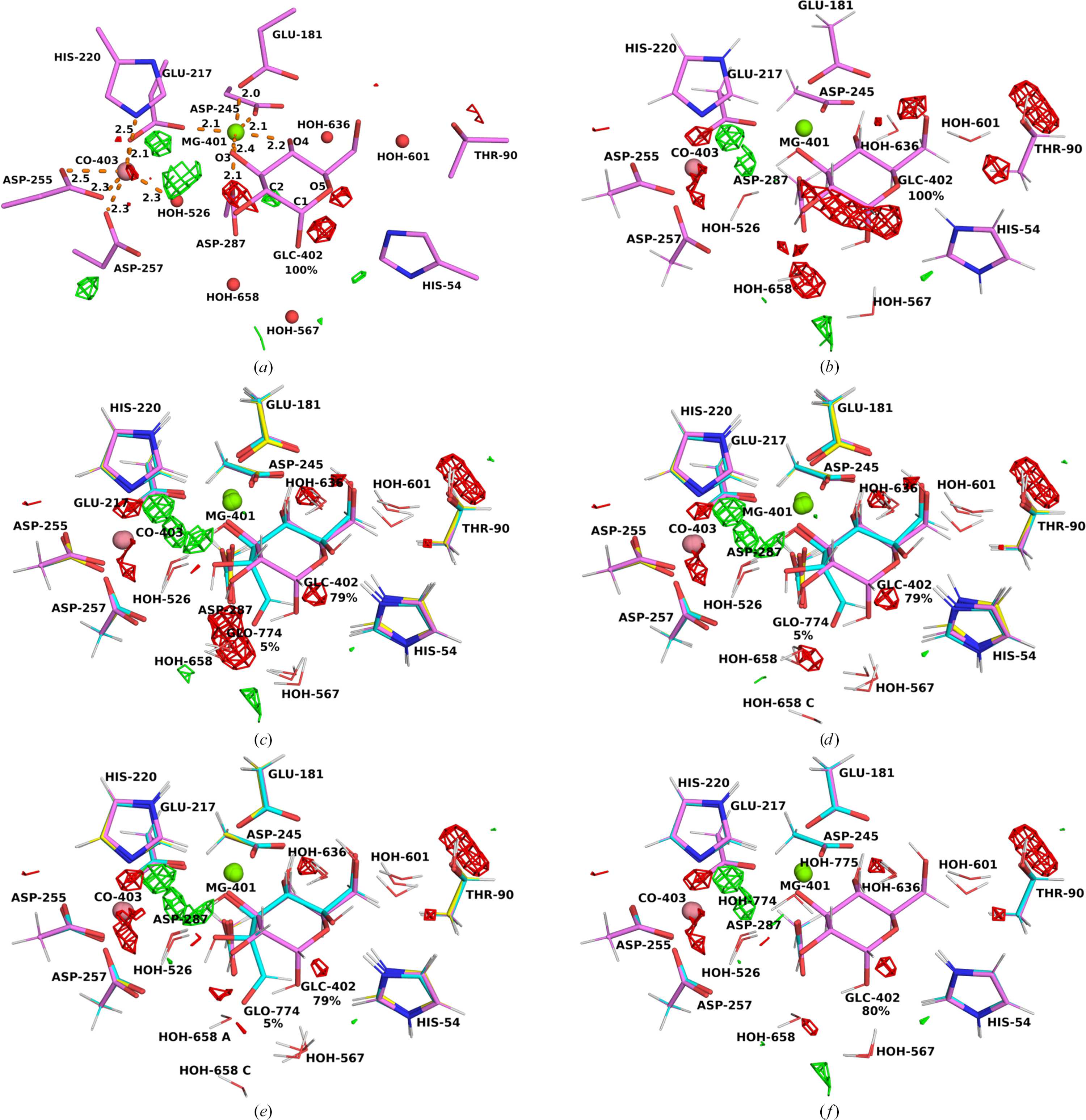
Figure 3
(a) Deposited structure of xylose isomerase (PDB entry 6qni). (b) QR structure with 100% Glc. (c) QR structure with 79% Glc, 5% Glo and 16% no substrate. (d) QR structure after movement of HOH658 in the 16% conformation. (e) QR structure after deleting HOH658 in the 5% conformation. (f) QR structure with 80% Glc and 20% no substrate. (b)–(f) illustrate the QM region used in QR. The conformation with Glc is depicted in pink, that with Glo in blue and that without substrate in yellow. The mFo − DFc difference maps are contoured at +3σ (green) and −3σ (red). |
IUCrJ
ISSN: 2052-2525
BIOLOGY | MEDICINE
Open  access