


 menu
|
|
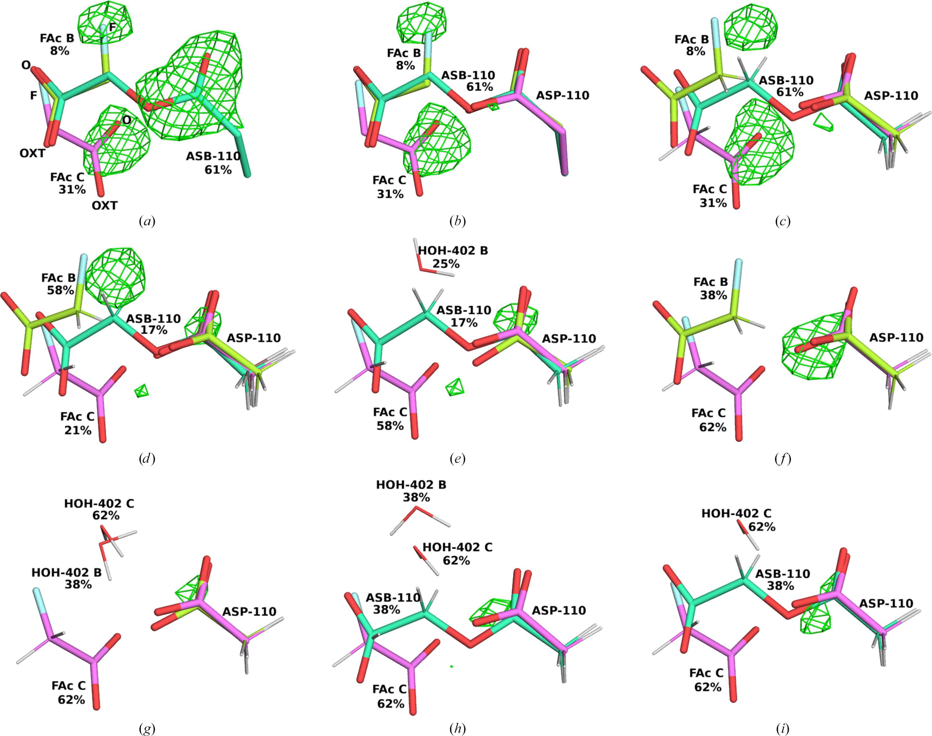
Figure 4
The deposited structure (PDB entry 6qhp) (a) and the various QR structures of the 2.3 s state of fluoroacetate dehalogenase. All structures show the active site of the A chain and are described in Table 1  . (c)–(i) illustrate the QM region used in QR. The different conformations are colour-coded with the C atoms in dark green, light green, magenta and brown, respectively. The mFo − DFc difference maps are contoured at +3σ (green; no significant negative differences are seen in the structures). |
IUCrJ
ISSN: 2052-2525
BIOLOGY | MEDICINE
Open  access