journal menu


issue contents
 | STRUCTURAL BIOLOGY |
ISSN: 2059-7983
Radiation Damage
Papers presented at the 12th International Workshop on X-ray Radiation Damage to Biological Crystalline Samples, 3–5 June 2025
Edited by Elspeth Garman and Martin Weik
This virtual issue collects together articles from the 12th International Workshop on X-ray Radiation Damage to Biological Crystalline Samples



Open  access
access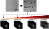
The effect of radiation damage in MicroED has been studied. By only including data collected at low accumulated electron dose during data processing, the effects of radiation damage can be reduced.

Open access
access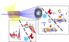
We used on-line in crystallo UV–Vis absorption spectroscopy in conjunction with X-ray crystallography on beamline BM07-FIP2 at the ESRF to compare the structural effects of specific radiation damage on two different metal-containing proteins at either room or cryogenic temperature.
PDB references: horse-heart myoglobin, room temperature, 32.5 kGy, 9t6v; 260.0 kGy, 9t6w; cryogenic temperature, 676.8 kGy, 9t6x; 14.4 kGy, 9t6y; nitrite reductase from Achromobacter cycloclastes, room temperature, 14.9 kGy, 9t6o; 283.1 kGy, 9t6p; cryogenic temperature, 33.3 kGy, 9t6q; 1332 kGy, 9t6u
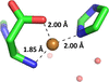
X-ray exposure influences the structures of Cu(I), Cu(II) and Zn(II) complexes of amyloid-β (Aβ) peptides in different ways. While Cu(I) and Zn(II) complexes show no detectable X-ray-induced spectral changes, Cu(II) complexes undergo significant spectral evolution under irradiation. Experiments on the truncated fragments Aβ1–6 and Aβ1–16 show that the extent of relaxation depends on the peptide length, on the binding mode and on the temperature.
Open access
access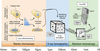
Cryo-EM analysis of apoferritin exposed to an X-ray dose of 100 MGy confirms the potential for high-resolution correlative imaging workflows combining X-ray tomography and cryo-EM.
Open access
access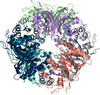
We characterize the formation of a ferryl state of a dye-decolourizing peroxidase by drop-on-chip serial femtosecond crystallography and also the modulation of the heme redox state using dose-resolved serial synchrotron crystallography.
Open access
access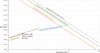
The use of X-rays and electrons for diffraction and imaging of soft biological samples is compared.
Open access
access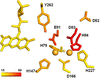
Flavodiiron proteins (FDPs) are di-iron enzymes that reduce NO and/or O2 with distinct substrate selectivity. We have produced E. coli FDP variants targeting second coordination-sphere residues and determined their structures. Although the kinetics remained unchanged, the E. coli FDP S262Y variant showed X-ray radiation-induced photoreduction, suggesting increased structural sensitivity without altered selectivity.
PDB references: Escherichia coli FDP, D52K mutant, oxidized state, 7r0f; D52K mutant, reduced state, 7r1h; S262Y mutant, oxidized state, 7r2o; S262Y mutant, reduced state, 7r1j; D52K/S262Y mutant, oxidized state, 7r2p; D52K/S262Y mutant, reduced state, 7r2r; D52K mutant, pressurized with krypton gas, 7r2s
Open access
access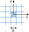
Analytical expressions for the damage-limited resolution are developed and applied to X-ray and electron imaging of beam-sensitive specimens. The findings will guide future microscopy and instrument design.
Open access
access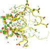
This first systematic radiation-damage study at 0.55 Å resolution reveals extensive small conformational changes in response to dose. The findings demonstrate the benefits of using large crystals fully bathed in a large `top-hat' beam with a uniform fluence profile, together with low-dose data-collection protocols, for sub-Ångström structure investigations on macromolecules.
Open access
access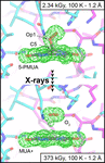
This manuscript investigates the X-ray-induced radiolysis of the catalytic C5-peroxide adduct in urate oxidase crystals, utilizing a top-hat X-ray beam to monitor peroxide occupancy across extensive dose series at both 100 K and room temperature. The study reveals a kinetic phase transition where the peroxide decays rapidly via zero-order kinetics at room temperature, whereas at 100 K the decay is retarded and follows first-order kinetics due to an efficient recombination mechanism involving trapped oxygen.
Open access
access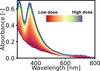
Radiation-induced changes during X-ray crystallographic data collection can compromise concurrent spectroscopic studies. Suitable protocols to detect and identify spectral artefacts from crystallization buffers and cryoprotectants are essential for the reliable analysis of chromophoric ligands in macromolecules.
Open access
access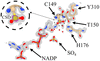
This work examines data contained within 118 215 structures in the Protein Data Bank for unmodelled difference density concomitant with unmodelled oxidatively damaged cysteines. This analysis demonstrates that site-specific oxidation of residues is a phenomenon of which the experimenter should be aware, and highlights the need for a suitable detection tool within refinement software.
Open access
access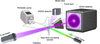
Adenylate kinase was used to quantify how experimental parameters influence radiation damage during time-resolved X-ray solution scattering (TR-XSS) at a multipurpose SAXS beamline. The study provides general guidelines for minimizing radiation artifacts in TR-XSS.
Open access
access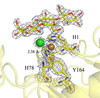
Small but measurable changes in the coordination geometry of a lytic polysaccharide monooxygenase copper active site are revealed by X-ray crystallography, providing structural insight into their catalytic flexibility.
PDB references: LsAA9A, cryo temperature, low-dose structures of Cu(II) state, 9tcx; 9tcv; 9tcy; low-dose structures of Cu(II) state with cellotriose, 9tcz; 9td0; 9td1; low-dose structures of Cu(I) state, 9td6; 9td8; 9td9; low-dose structures of Cu(I) state with cellotriose, 9tdb; 9tdc; 9tdd; high-dose structures of Cu(I) state, 9tdh; 9tdi; high-dose structures of Cu(I) state with cellotriose, 9tdj; 9tdk; room temperature, multicrystal structure at 1.5 kGy, 9tde; at 88.5 kGy, 9tdf

Download citation


Download citation
Open access
access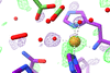
A series of 36 structures of the Neurospora crassa LPMO NcAA9D were refined against X-ray crystal datasets collected with increasing dose from a single crystal to provide insights into dose-dependent structural changes at the active site of the enzyme. The findings underscore the importance of minimizing accumulated dose during cryo-X-ray crystallographic studies of lytic polysaccharide monooxygenases to prevent the misinterpretation of radiation damage-related electron-density features.