

 journal menu


issue contents
January 2015 issue
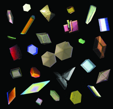
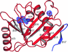
Cover illustration: Crystals on the cover. A selection of crystals from articles published in 2014.
crystals on the cover


Free 

IYCr crystallization series
Open access
access
A comprehensive and up-to-date review of the lipid cubic phase or in meso method for crystallizing membrane and soluble proteins and complexes is reported. Recent applications of the method for in situ serial crystallography at X-ray free-electron lasers and synchrotrons are described.
research communications


The Cu2+ ion of the blue copper protein pseudoazurin from A. faecalis was replaced by a Zn2+ ion and the resulting protein was phased by SAD using the anomalous signal of the S and Zn atoms. The Zn2+ ion lies closer to the plane of its strong ligand atoms (His40 Nδ1, Cys78 Sγ and His81 Nδ1) and attracts its axial ligand Met86 Sδ closer.
PDB reference: Zn-substituted pseudoazurin, 4rh4
The HU protein from the parasitic mycoplasma S. melliferum KC3 was cloned, overexpressed in E. coli and purified. Crystals of the protein that diffracted to 1.36 Å resolution were obtained for the first time.
L-Arabinose isomerase from L. fermentum CGMCC2921 was heterologously expressed, purified and crystallized, and preliminary X-ray diffraction analysis was successfully carried out.
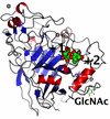
Crystal structures of Rab2 and Rab3 from D. melanogaster in complex with GMPPNP are presented.
A thermostable glycoside hydrolase family 116 β-glucosidase from T. xylanolyticum was expressed in E. coli, purified and crystallized. The crystal diffracted to 3.15 Å resolution with P21 symmetry.
A novel carbohydrate-binding module (CBM-Rf6A) of a modular family 9 glycoside hydrolase from R. flavefaciens was purified and crystallized, and data were collected for the native protein and a selenomethionine derivative to 1.75 and 1.5 Å resolution, respectively. The crystals belonged to orthorhombic and cubic space groups, respectively, and the structure was solved by a single-wavelength anomalous dispersion experiment.

DR0248, a MNT–HEPN fused protein from D. radiodurans, was expressed, purified and crystallized in two crystal forms. MAD phases were obtained from zinc identified in the protein by X-ray fluorescence.
This article describes the preliminary crystallographic data of SF173 from Shigella flexneri.

The type IV restriction endonuclease ScoMcrA, which cleaves both Dcm-methylated DNA and phosphorothioated DNA, was crystallized and the crystals diffracted to 3.35 Å resolution. The space group and the unit-cell parameters were determined.

The type I signal peptidase from S. aureus Newman strain was crystallized as a fusion protein with maltose-binding protein. Preliminary X-ray data showed that the crystals diffracted to 2.05 Å resolution.
The three-dimensional single-crystal X-ray structure of a thermally stable and industrially relevant bacterial α-amylase variant has been determined at 1.9 Å resolution.
PDB reference: engineeered bacterial α-amylase, 4uzu
Crystals of the C-terminal part of PorM obtained by limited proteolysis of the purified recombinant protein and grown from PEG solutions were tetragonal (space group P43212) and diffracted to 2.85 Å resolution.
Open access
access
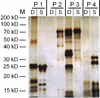
Application of in situ dynamic light scattering to solutions of protein–detergent complexes permits characterization of these complexes in samples as small as 2 µl in volume.

The CARD domain of apoptosis repressor with CARD (ARC) was purified and crystallized. Crystals were obtained at 293 K and diffracted to a resolution of 2.1 Å; they belonged to space group P61 or P65, with unit-cell parameters a = 98.28, b = 98.28, c = 51.86 Å, α = 90, β = 90, γ = 120°.
Structures of the catalytic N-acetyltransferase (NAT) domain of the bifunctional N-acetyl-L-glutamate synthase/kinase (NAGS/K) from Xylella fastidiosa bound to N-acetyl-L-glutamate (NAG) with and without an N-terminal His tag have been solved and refined at 1.7 and 1.4 Å resolution, respectively.

An NAD+-reducing [NiFe] hydrogenase was purified and crystallized. An X-ray diffraction data set was collected to a resolution of 2.58 Å.
A protease in the common commercial enzyme cleaner Zymit used for the cleaning of crystallization robots cannot be completely inactivated by EDTA.
SpaD, a backbone-pilin fragment from L. rhamnosus GG, was crystallized and X-ray diffraction data were collected to a resolution of 2.0 Å. In-house iodide-derivative data were collected to 2.2 Å resolution for phase determination by the single-wavelength anomalous dispersion method.
The cloning, expression, purification, crystallization and preliminary X-ray diffraction analysis of Rv3899c from M. tuberculosis are described.
A periplasmic sensory domain of the C. jejuni chemoreceptor for aspartate A (CcaA) has been purified and crystallized by the hanging-drop vapour-diffusion method. A diffraction data set has been collected to 1.4 Å resolution.
The structure of an industrially important thermally stable cellulase, A. fumigatus cellobiohydrolase I, has been solved in apo and product-complex forms at 1.8 and 1.5 Å resolution, respectively.
![[publBio]](/logos/publbio.gif)


