
Acta Crystallographica
Section B STRUCTURAL SCIENCE, CRYSTAL ENGINEERING AND MATERIALS


 menu


research papers
| STRUCTURAL SCIENCE CRYSTAL ENGINEERING MATERIALS |
 access
Sheared structure Pb4Sr14Fe23O52 solved by three-dimensional electron diffraction combined with atomic scale STEM imaging

aUniversity of Grenoble Alpes, CNRS, Grenoble INP, Institut Néel, 38000 Grenoble, France
*Correspondence e-mail: [email protected]
Ferrite Pb4Sr14Fe23O52 was synthesized in 2006, and its structure was partially described from the interpretation of high-resolution images. Twenty years later, thanks to the constant development in electron crystallography, the of this oxide is fully determined by combining three-dimensional electron diffraction with aberration-corrected (STEM). The compound crystallizes in the monoclinic lattice, C2/c, with unit-cell parameters a = 34.782 (8) Å, b = 5.522 (2) Å, c = 24.962 (7) Å and β = 99.11 (2)°. The dynamically refined structure consists of the collapse of a single perovskite layer and an oxygen-deficient rock-salt-type layer. This work demonstrates the potential of combining electron crystallography and atomic resolution STEM imaging to solve complex oxide structures that remained inaccessible using earlier experimental approaches.
Keywords: crystal structure; 3D electron diffraction; HAADF and ABF STEM; ferrite.
CCDC reference: 2585591
1. Introduction
Over the past fifteen years, three-dimensional electron diffraction (3D ED) has evolved into a powerful and routinely used technique for ab initio determination (Gemmi et al., 2019 ). This progress has been driven by major advances in electron microscopy instrumentation, including the development of fast and highly sensitive detectors, automated data acquisition protocols, and dedicated software for data processing and structure solution. The most commonly used programs are PETS2 for the processing of precession ED tomography and continuous rotation ED data (Palatinus et al., 2019), XDS for indexing, integration and scaling of diffraction intensities (Kabsch, 2010), RED for automated three-dimensional reconstruction and data processing (Wan et al., 2013) and DIALS, which provides a robust framework for the processing of ED data, particularly in MicroED and serial electron crystallography applications (Clabbers et al., 2018). These developments have greatly improved the accuracy, reliability and accessibility of 3D ED, enabling routine structure determination from crystals too small for conventional single-crystal X-ray diffraction. As a result, crystal structures that were previously inaccessible can now be routinely determined from submicrometric crystals. The capabilities of 3D ED have been demonstrated for a wide range of structurally complex materials, including zeolites (Örs et al., 2024; Li et al., 2024; Tirado et al., 2025a; Tirado et al., 2025b), metal–organic frameworks (Huang et al., 2021; Gorelik et al., 2026), and both commensurately and incommensurately modulated structures (Mei et al., 2023; Plana-Ruiz et al., 2024).
Before the widespread adoption of automated 3D ED methods, ED data had already been used for quantitative crystal structure analysis, including structure solution and refinement from two-dimensional diffraction patterns. Several approaches have been developed to extract structural information from such data, including direct methods and least-squares refinement procedures using electron scattering factors. In particular, the MSLS (MultiSlice Least-Squares) approach enabled quantitative structure refinement from dynamical ED patterns by explicitly taking multiple scattering into account (Jansen et al., 1998). Structure solution and refinement from ED data had therefore already been demonstrated before the emergence of modern 3D ED workflows. The main advance brought by modern 3D ED was not the possibility of extracting structural information from ED itself, but rather the development of experimental and computational workflows that allow substantially more complete three-dimensional reciprocal space datasets to be collected and processed in a systematic and increasingly automated manner. Earlier approaches were often based on a limited number of two-dimensional diffraction patterns recorded along selected zone axes and could require more specialized data collection, processing and refinement strategies. In addition, the strong influence of dynamical scattering on ED intensities could complicate quantitative structure analysis, particularly for thicker crystals. Nevertheless, dynamical refinement methods had already demonstrated that reliable structural information could be extracted from ED data when multiple scattering was explicitly considered (Jansen et al., 1998). The subsequent development of automated 3D ED acquisition and processing has made such analyses considerably more accessible and has facilitated the routine determination of increasingly complex crystal structures. However, the structural complexity of some compounds may require the combination of single-crystal X-ray diffraction and ED to achieve a reliable structure determination. A representative example is the complex incommensurately modulated KNd(MoO4)2 (Zhou et al., 2016). Moreover, because electrons interact with the electrostatic potential rather than the electron density, ED is particularly sensitive to light atoms, making it possible to locate O and even H atoms that are often difficult to detect by conventional X-ray diffraction (Palatinus et al., 2017; Klar et al., 2023; Yörük et al., 2025). Consequently, numerous compounds whose structures could not previously be solved because of crystal size limitations or structural complexity have recently become accessible to complete crystallographic characterization.
In this context of complex structures, the layered ferrites belonging to the homologous series [Sr2Fe4O7–δ][Sr2Fe2O6]m, consist of the periodic intergrowth of one oxygen-deficient rock-salt-type layer and m perovskite layers. The parent member (m = 1), Sr4Fe6O13–δ [Fig. 1(a)] exhibits an incommensurately modulated structure originating from the distribution of oxygen vacancies within the rock-salt-type layer. The modulated feature of this structure was identified by single-crystal X-ray diffraction study, which showed three different environments of Fe in the oxygen-deficient rock-salt-type layer: trigonal bipyramid (TBP), tetragonal pyramid (TP) and tetrahedron (T) (Mellenne et al., 2004; Pérez et al., 2006). Partial substitution of Sr2+ by stereochemically active cations such as Bi3+ or Tl+ stabilizes the m = 2 [Fig. 1(b)] and m = 4 [Fig. 1(c)] members of the series, corresponding to the compositions (BiSr3.5)Fe6O14 (Lepoittevin et al., 2004; Grebille et al., 2006) and (TlɛSr5–ɛ)Fe6O15.4 (Lepoittevin, 2016). These two structures were solved using single-crystal X-ray diffraction and 3D ED, respectively, and in contrast to the parent phase, they exhibit commensurate modulation that can be described using well defined superstructures. Motivated by the possibility of stabilizing new members of this family through cation substitution, the solid solution (Sr4–xPbx)Fe6O13±δ was subsequently investigated (Lepoittevin et al., 2006). For x = 0.7, a previously unknown phase was isolated and identified as a new lead-substituted ferrite. At the time, the complete crystallographic characterization of this complex structure from ED remained challenging because of the limited coverage provided by two-dimensional diffraction data and the need to account for dynamical scattering. A preliminary structural model, established from high-resolution (HRTEM), selected area ED, and energy-dispersive X-ray (EDX) spectroscopy, indicated that this phase originated from the shearing of a single perovskite layer and an oxygen-deficient rock-salt-type layer [Fig. 1(d)]. Although this study successfully described the overall structural framework, essential crystallographic information remained inaccessible, particularly the distribution of the Pb atoms and the arrangement of O atoms within the oxygen-deficient rock-salt-type layer.
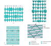 | Figure 1 Structures of (a) Sr4Fe6O13–δ, (b) (BiSr3.5)Fe6O14, (c) (TlɛSr5–ɛ)Fe6O15.4 and (d) first structural model of so-called `Pb4Sr13Fe24O53' deduced by zone axis ED and HRTEM images interpretation in 2006. |
In the present work, we revisit this compound using the current state of the art electron crystallography toolbox. By combining ab initio structure solution from 3D ED with aberration-corrected HAADF and annular bright-field (ABF) STEM imaging and dynamical we establish the complete of Pb4Sr14Fe23O52.
2. Experimental
2.1. Synthesis
The single-phase powder was synthesized under an inert atmosphere in a using PbO, SrO, SrO2 and Fe2O3 as starting materials. SrO was obtained by thermal decomposition of Sr(OH)2·8H2O at 1100 °C and subsequently maintained at this temperature until use to prevent hydration. The precursor powders were weighed according to the nominal stoichiometric composition and thoroughly homogenized by grinding in an agate mortar. The resulting powder mixture was uniaxially pressed into bar-shaped pellets, sealed in an evacuated silica tube, and subjected to a heat treatment at 1100 °C for 48 h. The heating and cooling steps were both performed at a controlled rate of 1.5 °C min−1.
2.2. Transmission electron microscopy (TEM)
The TEM specimen was prepared by dispersing a small amount of powder in ethanol and depositing a drop of the suspension onto a holey carbon-coated copper grid. 3D ED and STEM analyses were performed on a probe-corrected Jeol NEOARM 200F microscope operating at 200 kV, equipped with a Nanomegas Digistar precession device and a 4K CMOS Gatan OneView camera. Atomic scale chemical contrast imaging was performed using high-angle annular dark field (HAADF) and ABF detectors in STEM mode. Stepwise ED tomography data were collected using a ±70° tomography specimen holder, using 1° increments and a 1.2° precession angle to ensure full integration of diffracted intensities (Mugnaioli et al., 2009). Data analysis and 3D reciprocal space reconstruction were conducted in PETS2 (Process Electron Tilt Series, version 2) software (Palatinus et al., 2019). The structural model was calculated using the charge flipping algorithm (Palatinus, 2013) implemented in Superflip (Palatinus & Chapuis, 2007) and subsequently kinematically and dynamically refined in JANA2020 (Petříček et al., 2023).
3. Results
3.1. Unit-cell and structural model determination
A 3D ED dataset was collected on an isolated single crystallite selected to avoid any overlap with neighbouring particles throughout the entire tilt series. Prior to diffraction data acquisition, the crystal composition was assessed using EDX, confirming the presence of Pb, Sr and Fe with approximate atomic proportions of 10%, 40% and 50%, respectively. These values are consistent with the targeted Pb–Sr ferrite phase and were used to verify the chemical of the selected crystal. The diffraction dataset was acquired over an angular range from +60° to −60°. To ensure that diffraction intensities originated from the same crystal volume during the complete rotation experiment, a 20 µm selected area aperture was employed, allowing the illuminated region to remain confined to the same area of the crystallite throughout the data collection. The 121 ED frames were processed using PETS2 software. Diffraction spots were first automatically detected and indexed throughout the tilt series. From these data, PETS2 determined the experimental rotation axis, giving rise to the sinogram shown in Fig. 2(a). The software then reconstructed the three-dimensional reciprocal space, enabling the determination of the unit-cell parameters and the crystal orientation, which was subsequently refined during the data reduction procedure. The refined unit cell was found to be monoclinic, with the unit-cell parameters a = 34.782 (8) Å, b = 5.522 (2) Å, c = 24.962 (7) Å and β = 99.11 (2)°. Using the resulting orientation matrix, reflection intensities were extracted from each diffraction frame. During the data reduction procedure, PETS2 performs several refinements and corrections to improve the internal consistency of the dataset. These include of the beam centres on individual diffraction patterns, correction of the experimental tilt angles, compensation for geometrical distortions of the diffraction frames, and of both the orientation matrix and the unit-cell parameters. The quality of the processed data was assessed through the rocking curve representation, commonly called the camel plot [Fig. 2(b)]. This plot provides a graphical evaluation of the reflection intensity profiles and constitutes a reliable indicator of the quality of the dataset for subsequent ab initio and structure dynamical refinement.
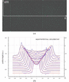 | Figure 2 (a) Sinogram representing s(θ) (reflection position) versus θ (rotation angle) and (b) the camel plot after PETS2 processing. |
Selected sections of the were calculated in order to analyse the and identify the compatible space groups. These sections, depicted in Fig. 3, reveal the systematic extinction condition h + k = 2n for the general reflections hkl, which is consistent with a C centering. In addition, the h0l section exhibits the extinction condition l = 2n, indicating the presence of a c glide mirror perpendicular to the b axis. These conditions limiting the reflections are compatible with the monoclinic space groups C2/c and Cc. Following the final integration of the diffraction intensities, PETS2 generated two reflection files: a conventional hkl file for ab initio structure resolution and a second for subsequent dynamical During the data reduction procedure, several were evaluated, and the monoclinic 2/m symmetry was retained as it yielded the lowest merging residual Rint.
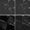 | Figure 3 Selected sections of the reciprocal space exhibiting reflections indexing consistent with the space groups C2/c and Cc. |
The structural model was calculated from the integrated reflection intensities exported by PETS2. The dataset used for the structure solution comprised 6862 independent reflections, corresponding to a completeness of 87% up to a resolution of 0.70 Å. The charge flipping calculation successfully converged toward an initial electrostatic potential distribution, providing a first structural model of the investigated Pb–Sr ferrite. The solution was obtained with an agreement factor of 30.71% in space group C2/c.
The resulting structural model presented in Fig. 4(a) closely resembles the first one reported in 2006. It consists of a framework built up from the collapse of single perovskite layers alternating with rock-salt-type oxygen-deficient double iron layers. The main features of the framework are therefore correctly reproduced by the charge flipping solution, confirming the validity of the structural model derived from the 3D ED data. However, this first model remains incomplete: several O atoms are absent, particularly within the oxygen-deficient double iron layers. Furthermore, the Pb and Sr cations appear to be randomly distributed over the same crystallographic sites, preventing an unambiguous assignment of their occupancies at this stage of the structure determination. This observation can be explained by comparison of electron and X-ray scattering factors of Pb, Sr and Fe over the same scattering vector range 0 ≤ Q (Å−1) ≤ 0.7. Based on values from Doyle & Turner (1968), X-ray scattering factors are, to a first approximation, proportional to the atomic number Z. Consequently, the large difference between Z(Pb) = 82 and Z(Sr) = 38 results in a pronounced separation of their X-ray scattering factors [Fig. 5(a)], making the two elements readily distinguishable. On the other hand, the difference between their electron scattering factors is much less pronounced over the same Q range [Fig. 5(b)]. The distinction between Pb and Sr is more directly visualized in HAADF-STEM images, owing to a strong dependence on atomic number Z. In order to search for the information of Pb/Sr and O atoms distribution in the structure, atomic scale STEM imaging was then employed.
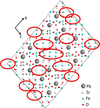 | Figure 4 Raw structural model calculated by Superflip. The regions circled in red correspond to missing O atoms. |
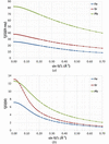 | Figure 5 (a) X-ray and (b) electron scattering factor curves of Fe, Sr and Pb atoms plotted using values from Doyle & Turner (1968 ). |
3.2. HAADF and ABF STEM
HAADF and ABF STEM provide complementary structural information. In HAADF STEM imaging, the contrast is approximately proportional to Zn with n ≃ 2 (Pennycook et al., 2000), producing a contrast in which heavy elements appear significantly brighter than lighter ones. This imaging mode is particularly suitable for identifying the distribution of heavy cations such as Pb (Z = 82), Sr (Z = 38) and Fe (Z = 26). In contrast, ABF STEM collects electrons scattered at lower angles, and the image contrast arises from the interference between the transmitted and diffracted electron beams. Consequently, the contrast does not follow a simple dependence on the but is also influenced by factors such as specimen thickness, crystal orientation and defocus. Under suitable imaging conditions, ABF STEM enables the direct visualization of light element atomic columns, such as oxygen.
Fig. 6(a) exhibits an HAADF STEM image acquired along the [010] zone axis. On this image, the Pb atomic columns are clearly identified as the brightest dots. They appear as groups of four atomic columns arranged in a characteristic staircase like configuration, directly revealing the displacement of the perovskite blocks responsible for the sheared nature of the structure. This observation provides direct experimental evidence that the Pb cations are intimately associated with the collapse mechanism. Such a collapse of layered frameworks induced by the incorporation of stereochemically active heavy cations is not unprecedented and has been previously reported in several complex oxides containing Bi3+ or Pb2+. In particular, Bi-based layered oxides have been shown to undergo shear-induced structural rearrangements leading to sheared structures, where the stereochemical activity of the Bi 6s2 lone pair promotes distortions of the interlayer regions and stabilizes displaced perovskite blocks (Hervieu et al., 1995; Hervieu et al., 1997). The image also shows dots with slightly lower intensity assigned to Sr. Between the Sr layers, the Fe atoms are also clearly visible, despite their weaker contrast. The iron sublattice is organized into two distinct structural motifs: single rows composed of 11 Fe atomic columns and oxygen-deficient double rows systematically consisting of six pairs of Fe columns. These two arrangements alternate throughout the structure and are consistent with the layered framework deduced from structure solution, in which perovskite-like slabs alternate with oxygen-deficient rock-salt-type double Fe layers.
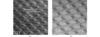 | Figure 6 (a) [010]-oriented HAADF STEM image. White circles highlight Pb atomic columns. (b) [010]-oriented ABF STEM image, where oxygen atomic columns are pointed out by white arrows. |
The [010] ABF STEM image provides complementary information on the oxygen [Fig. 6(b)]. Although the oxygen atomic columns exhibit a weak contrast barely distinguishable from the background, they can nevertheless be identified in both the single and double iron layers. Within the single Fe layers, O atoms are observed between Fe atoms. In double iron layers, pairs of oxygen atoms are adjacent to four pairs of Fe atoms, whereas only a single oxygen column is observed on each side of the double iron layer. All oxygen positions already present in the calculated structural model are consistent with the contrast observed in the ABF STEM image. ABF observations also offer valuable guidance for locating the O atoms that remain absent from the structural model. In the subsequent refinement procedure, these observations will be systematically compared with difference fast Fourier transform (FFT) maps calculated after each refinement cycle, allowing the missing oxygen sites to be identified and incorporated into the model.
3.3. Structure refinement
Based on the information obtained from the HAADF STEM image, the Pb atomic positions were reassigned to match the experimentally observed cation distribution. In particular, the staircase-like arrangement of Pb columns, directly visualized by Z contrast imaging, enabled an unambiguous distinction between Pb and Sr crystallographic sites. A kinematical of the modified structural model was subsequently performed. This served as the basis for the calculation of difference FFT maps in order to reveal positive residual electrostatic potential and locate missing atomic positions, particularly those corresponding to O atoms. As shown in Fig. 7, numerous positive residual maxima are observed in regions where O atoms are expected according to the ABF STEM observations. The agreement between these residual electrostatic potentials and the oxygen columns identified in the ABF confirm that these maxima correspond to missing oxygen sites. Consequently, O atoms were progressively introduced at these positions, leading to a more complete structural model.
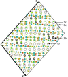 | Figure 7 Difference FFT map created after kinematical refinement. Residual positive electrostatic potentials (yellow dots) reveal the positions of the last missing O atoms in the structure. |
After having completed the structural model, a full was carried out using the of ED (Palatinus et al., 2015). In contrast to the kinematical approximation, the dynamical refinement accounts for multiple scattering effects and precession motion, providing a significantly more accurate description of the interaction between the electron beam and the crystal. As expected, this resulted in a substantial improvement of the agreement between calculated and experimental diffraction intensities. In particular, the observed reliability factor, Robs, was reduced by approximately a factor of 2.5 compared with the value obtained from the kinematical refinement. To assess the completeness of the final structural model, a last difference FFT map was calculated after convergence of the dynamical refinement. The remaining residual peaks are attributed to background noise, as they exhibit insufficient intensity to justify the introduction of new atoms [Fig. S1(a)]. Furthermore, several of these weak maxima are located at chemically unrealistic distances from the atoms already present in the refined structure, excluding their interpretation as physically meaningful atomic sites [Fig. S1(b)]. The absence of significant residual features therefore confirms that the final structural model provides a complete and self-consistent description of the crystal structure. The crystallographic parameters of the final structural model, including the refined unit-cell parameters, space group, atomic coordinates and atomic displacement parameters are summarized in Table 1. The reliability factors together with the parameters of the dynamical refinement are also reported. Slightly negative ADPs are obtained for O5, O9, O18 and O24. These values should not be interpreted as physically meaningful negative atomic displacements, but rather as an indication that the corresponding displacement parameters are poorly defined within the refinement. Their proximity close to zero probably reflects the limited precision with which the ADPs of light atoms can be determined from 3D ED data. Similar anomalous ADPs, including negative values, have been reported in 3D ED refinements and were shown to be sensitive to the quality and processing of the ED data (Yang et al., 2022).
| ||||||||||||||||||||||||||||||||||||||||||||||||||||||||||||||||||||||||||||||||||||||||||||||||||||||||||||||||||||||||||||||||||||||||||||||||||||||||||||||||||||||||||||||||||||||||||||||||||||||||||||||||||||||||||||||||||||||||||||||||||||||||||||||||||||||||||||||||||||||||||||||||||||||||||||||||||||||||||||||||||||||||||||||||||||||||||||||||||||||||||||||||
4. Final structure and discussion
4.1. Structure description
The final structure, depicted in Fig. 8, corresponds to the chemical composition Pb4Sr14Fe23O52 and consists of a periodic alternation of two distinct structural blocks. The first block is a perovskite-like layer containing nine Fe atoms, all in octahedral environment. This slab alternates with an oxygen-deficient rock-salt-type layer composed of six pairs of Fe atoms exhibiting a variety of coordination environments arising from the ordered distribution of oxygen vacancies. Within this layer, the Fe atoms are found in tetrahedral (T), tetragonal pyramidal (TP) and trigonal bipyramidal (TBP) environments, following the ordered sequence TP-T-TBP-TBP-T-TP. The main feature of this structure is the periodic collapse of these two slabs, directly associated with the incorporation of Pb atoms. As shown by HAADF STEM imaging and confirmed by the dynamical a block of four Pb atoms adopt a characteristic staircase-like arrangement, producing a displacement of  +
 of adjacent Fe single layers, giving rise to the sheared structure. The stereochemical influence of Pb is reflected in the local coordination of the neighbouring Fe atoms, since those immediately above and below a pair of Pb systematically adopt a square pyramidal environment. The representation of the different Fe environments with the corresponding Fe–O distances is displayed in Fig. 9. To further assess the local coordination environments of the Fe atoms, bond valence sum (BVS) calculations were performed using the final refined structural model. The BVS method estimates the oxidation state of each Fe site from its coordination number and the corresponding Fe–O bond distances. The bond valence sij was calculated according to the expression proposed by Brown & Altermatt (1985):
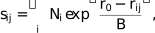
where Ni is rij is the metal–ligand distance, r0 and B are empirical parameters. Values of r0 and B used for our calculations are the ones reported for Fe–O bonds by Kanowitz & Palenik (1998). The estimated BVSs for the individual Fe sites are summarized in Table 2. Overall, the calculated values indicate that the majority of the Fe atoms exhibit an oxidation state around Fe3+, except for Fe8, for which the calculated BVS is closer to 2, indicating an oxidation state approaching Fe2+. This site corresponds to the Fe atom located in a square pyramidal coordination environment, immediately above and below a pair of Pb atoms. By considering these oxidation states for Fe, as well as conventional +2 for Pb/Sr and −2 for O, the charge neutrality is yielded in agreement with the chemical composition Pb4Sr14Fe23O52.
| ||||||||||||||||||||||||||||||||||||||
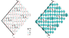 | Figure 8 Refined structure projected along [010] direction (a) without and (b) with Fe coordination polyhedra. |
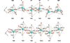 | Figure 9 Representation of the Fe environments and Fe–O distances in the (a) oxygen-deficient rock-salt-type layer (TP = tetragonal pyramid, TBP = trigonal bipyramid and T = tetrahedron) and (b) perovskite-type layer. |
Another feature of the structure is the corrugation of the layers. Rather than being perfectly planar, both the perovskite and oxygen-deficient rock-salt-type layers exhibit a periodic undulation throughout the structure. This distortion originates from the ordered oxygen distribution within the rock-salt-type layer. The central part of the layer, with the tetragonal pyramids, is significantly larger than its outer regions, which contain tetrahedra and trigonal bipyramids. This geometrical variation is directly transmitted to the adjacent perovskite slabs, which accommodate the distortion by adopting the same periodic corrugation.
4.2. Discussion
The structure exhibits strong similarities with the parent compound Sr4Fe6O13–δ. As a reminder, this phase has been described as an intergrowth of a single perovskite layer with an oxygen-deficient rock-salt-type layer, giving rise to three distinct coordination environments for the Fe atoms within the oxygen-deficient block, namely tetragonal pyramidal (TP), tetrahedral (T) and trigonal bipyramidal (TBP) sites. In the parent compound, however, the ordering of oxygen vacancies is incommensurate with the underlying lattice, resulting in an aperiodic succession of these coordination polyhedra. The modulation of the structure therefore directly originates from the distribution of oxygen vacancies within the rock-salt-type layer. In contrast, the Pb-containing phase investigated in the present work exhibits a fully periodic arrangement of the oxygen vacancies, giving rise to three Fe coordination environments following the sequence TP-T-TBP-TBP-T-TP, which is repeated throughout the structure. The introduction of Pb involves a long-range periodic ordering of the oxygen vacancies, leading to a commensurate modulation of the crystal structure. Such a stabilization of a commensurate modulated phase has also been reported in other members of this family, notably in the m = 2 compound containing double perovskite layers substituted with Bi (Lepoittevin et al., 2004), and in the m = 4 compound containing quadruple perovskite layers doped with Tl (Lepoittevin, 2016).
A further structural comparison can be carried out with the Pb-containing ferrate Pb4Sr2Fe6O15 (Fig. S2), which also adopts an oxygen-deficient perovskite-related framework (Raynova-Schwarten et al., 1997). However, the way in which the oxygen-deficient and perovskite layers are arranged differs between the two structures. Pb4Sr2Fe6O15 can be described as a regular intergrowth along the c axis of a double perovskite layer [Sr2Fe2O6], with a single oxygen-deficient layer [Sr2Fe2O4]. Within the latter, oxygen vacancies give rise to edge-sharing FeO5 square pyramids, which form chains that are interconnected through FeO6 octahedra. In contrast, in Pb4Sr14Fe23O52, the presence of Pb induces a collapse of the two types of layers, resulting in a structural arrangement comprising an oxygen-deficient double Fe layer associated with a single perovskite-type layer. The ordered distribution of oxygen vacancies gives rise to three distinct Fe environments, namely tetragonal pyramidal, tetrahedral and trigonal bipyramidal, arranged in a periodic sequence. This collapse of the perovskite and oxygen-deficient layers modifies the stacking sequence and breaks the higher symmetry of the parent layered arrangement, thereby, giving rise to a monoclinic symmetry. Thus, although both structures can be understood in terms of intergrowth of oxygen-deficient and perovskite-type layers, the incorporation of Pb results in a fundamentally different periodic arrangement of these layers and of the oxygen vacancies.
5. Conclusion
In this study, the of the ferrite Pb4Sr14Fe23O52 synthesized in 2006 has been finally successfully solved by 3D ED combined with atomic resolution STEM imaging. The structural model refined using the consists of a sheared layered framework resulting from the ordered incorporation of Pb atoms. The structure consists of the periodic collapse of two distinct structural blocks: a single perovskite layer and an oxygen-deficient rock-salt-type layer. Within the latter, the ordered distribution of oxygen vacancies gives rise to a regular sequence of Fe coordination polyhedral (TP-T-TBP-TBP-T-TP) and induces the characteristic corrugation of the layered framework. Bond valence sum calculations further indicate that most Fe atoms are trivalent, whereas the square pyramidal Fe site located adjacent to the Pb atoms exhibits an close to Fe2+, highlighting the strong influence of the local environment created by the Pb-induced collapse. The structure exhibits similarities with the parent phase Sr4Fe6O13–δ, but in contrast to this one the modulation appears to be commensurate, revealing an ordering of the oxygen vacancies. This feature was already observed in the Bi- and Tl-doped compounds, suggesting that these cations play a key role in promoting long-range ordering of oxygen vacancies and superstructures stabilization. Comparison with Pb4Sr2Fe6O15 highlights the structural role of Pb, as the two compounds exhibit on the one hand distinct arrangements of the oxygen-deficient and perovskite layers, and on the other hand different oxygen vacancies ordering. This work finally shows the efficiency of combining 3D ED with aberration-corrected STEM imaging for the determination of complex oxide structures.
Supporting information
CCDC reference: 2585591
contains datablocks global, I. DOI: https://doi.org/10.1107/S2052520626009285/je5067sup1.cif
PETS2 data in format. DOI: https://doi.org/10.1107/S2052520626009285/je5067sup2.txt
Figs S1 and S2. DOI: https://doi.org/10.1107/S2052520626009285/je5067sup3.pdf
| Fe23O52Pb4Sr14 | Z = 4 |
| Mr = 4171.88 | F(000) = 7496 |
| Monoclinic, C2/c | Dx = 5.853 Mg m−3 |
| Hall symbol: -C 2yc | Electron radiation, λ = 0.02511 Å |
| a = 34.782 (8) Å | Cell parameters from 871 reflections |
| b = 5.5225 (16) Å | θ = 0.1–0.9° |
| c = 24.962 (7) Å | µ = 0 mm−1 |
| β = 99.11 (2)° | T = 293 K |
| V = 4734 (2) Å3 | Needle |
| JEOL NEO ARM microscope diffractometer | θmax = 0.9°, θmin = 0.1° |
| 62683 measured reflections | h = −43→43 |
| 8671 independent reflections | k = −6→6 |
| 1222 reflections with I > 3σ(I) | l = −30→30 |
| Refinement on F | 0 restraints |
| R[F2 > 2σ(F2)] = 0.120 | 0 constraints |
| wR(F2) = 0.122 | Primary atom site location: charge flipping |
| S = 1.56 | Weighting scheme based on measured s.u.'s w = 1/(σ2(F) + 0.01F2) |
| 8671 reflections | (Δ/σ)max = 0.099 |
| 318 parameters |
Refinement. Structure solved by charge flipping (SUPERFLIP) and refined dynamically against 3D electron diffraction (precession electron diffraction tomography) data with JANA2020. Because of the limited tilt range inherent to the electron-diffraction data collection, reciprocal space coverage and data completeness at high resolution are lower than typical for X-ray single-crystal studies (see responses to PLAT026/027/029 below). |
| x | y | z | Uiso*/Ueq | ||
| Pb1 | 0.2348 (2) | 0.4948 (18) | 0.2837 (2) | 0.103 (3) | |
| Pb2 | 0.29664 (11) | 0.9937 (18) | 0.3674 (2) | 0.049 (2) | |
| Sr1 | 0.35308 (15) | 0.513 (2) | 0.4540 (3) | 0.0121 (17)* | |
| Sr2 | 0.33975 (16) | 0.991 (2) | 0.6272 (3) | 0.0143 (17)* | |
| Sr3 | 0.46800 (16) | 1.509 (2) | 0.6221 (3) | 0.0173 (19)* | |
| Sr4 | 0.40960 (16) | 0.984 (2) | 0.5387 (3) | 0.0127 (18)* | |
| Sr5 | 0.52975 (16) | 0.9782 (19) | 0.7020 (2) | 0.0139 (17)* | |
| Sr6 | 0.2799 (2) | 0.4965 (17) | 0.5412 (4) | 0.0212 (13)* | |
| Sr7 | 0.40500 (16) | 0.506 (2) | 0.7128 (3) | 0.0159 (16)* | |
| Fe1 | 0.5107 (2) | 1.072 (3) | 0.5619 (4) | 0.021 (3)* | |
| Fe2 | 0.4554 (2) | 0.424 (3) | 0.4833 (3) | 0.015 (2)* | |
| Fe3 | 0.2593 (2) | 0.499 (3) | 0.4075 (3) | 0.011 (2)* | |
| Fe4 | 0.10239 (19) | 0.571 (2) | 0.1084 (3) | 0.0063 (18)* | |
| Fe5 | 0.4286 (2) | 0.572 (3) | 0.3555 (3) | 0.022 (2)* | |
| Fe6 | 0.3164 (2) | 1.003 (3) | 0.4957 (4) | 0.025 (3)* | |
| Fe7 | 0.4353 (2) | 0.999 (3) | 0.6679 (3) | 0.020 (2)* | |
| Fe8 | 0.1998 (2) | 1.004 (3) | 0.3200 (4) | 0.021 (2)* | |
| Fe9 | 0.13980 (19) | 0.477 (3) | 0.2330 (3) | 0.014 (2)* | |
| Fe10 | 0.3354 (3) | 0.466 (4) | 0.3015 (5) | 0.057 (3)* | |
| Fe11 | 0.3752 (2) | 0.495 (3) | 0.5865 (3) | 0.013 (2)* | |
| Fe12 | 0.5 | 0.508 (4) | 0.25 | 0.030 (4)* | |
| O1 | 0.3493 (6) | 0.280 (6) | 0.5376 (10) | 0.032 (8)* | |
| O2 | 0.2300 (6) | 0.739 (6) | 0.3518 (9) | 0.015 (6)* | |
| O3 | 0.1325 (5) | 0.668 (4) | 0.1701 (7) | 0.022 (5)* | |
| O4 | 0.3480 (5) | 0.751 (5) | 0.5419 (7) | 0.000 (5)* | |
| O5 | 0.4086 (4) | 1.255 (4) | 0.6319 (7) | −0.006 (4)* | |
| O6 | 0.1007 (5) | 0.519 (6) | 0.2700 (8) | 0.025 (5)* | |
| O7 | 0.2402 (7) | 0.254 (7) | 0.3492 (12) | 0.061 (10)* | |
| O8 | 0.1757 (5) | 0.252 (5) | 0.2629 (8) | 0.011 (5)* | |
| O9 | 0.2899 (4) | 1.260 (4) | 0.4470 (7) | −0.008 (4)* | |
| O10 | 0.4133 (4) | 0.403 (5) | 0.4103 (7) | 0.018 (5)* | |
| O11 | 0.2888 (6) | 0.763 (6) | 0.4474 (10) | 0.020 (7)* | |
| O12 | 0.5582 (4) | 1.116 (4) | 0.6177 (7) | 0.012 (4)* | |
| O13 | 0.4635 (7) | 1.098 (8) | 0.4932 (11) | 0.061 (8)* | |
| O14 | 0.5287 (7) | 1.232 (6) | 0.2870 (10) | 0.020 (5)* | |
| O15 | 0.5319 (6) | −0.247 (6) | 0.2974 (10) | 0.021 (5)* | |
| O16 | 0.5989 (5) | 1.231 (5) | 0.3799 (8) | 0.022 (6)* | |
| O17 | 0.5369 (6) | 0.497 (7) | 0.1893 (9) | 0.028 (6)* | |
| O18 | 0.3369 (4) | 0.456 (4) | 0.6310 (6) | −0.004 (3)* | |
| O19 | 0.2815 (5) | 0.981 (4) | 0.5500 (8) | 0.018 (4)* | |
| O20 | 0.3782 (6) | 0.368 (6) | 0.8149 (9) | 0.045 (7)* | |
| O21 | 0.4903 (6) | 0.420 (8) | 0.9469 (11) | 0.075 (9)* | |
| O25 | 0.6754 (8) | 0.762 (8) | 0.7628 (12) | 0.066 (10)* | |
| O27 | 0.5224 (5) | −0.015 (7) | 0.8986 (9) | 0.027 (6)* | |
| O22 | 0.5778 (3) | 0.534 (4) | 0.9694 (5) | −0.009 (3)* | |
| O23 | 0.1918 (3) | 1.021 (5) | 0.1469 (5) | 0.001 (3)* | |
| O26 | 0.1375 (5) | 0.503 (6) | 0.0628 (8) | 0.027 (6)* |
| U11 | U22 | U33 | U12 | U13 | U23 | |
| Pb1 | 0.216 (7) | 0.037 (6) | 0.048 (4) | −0.061 (7) | −0.003 (4) | 0.002 (4) |
| Pb2 | 0.0324 (16) | 0.075 (6) | 0.044 (3) | −0.014 (4) | 0.0175 (16) | 0.006 (4) |
| Pb1—O2 | 2.20 (3) | Sr7—O14viii | 2.73 (3) |
| Pb1—O7 | 2.09 (3) | Sr7—O15xvii | 2.66 (3) |
| Pb1—O8 | 2.44 (2) | Sr7—O16viii | 2.72 (2) |
| Pb1—O25i | 2.45 (3) | Sr7—O17x | 2.92 (2) |
| Pb1—O23ii | 2.853 (13) | Sr7—O18 | 2.883 (13) |
| Pb2—O2 | 2.69 (2) | Sr7—O20 | 2.95 (3) |
| Pb2—O7iii | 2.42 (3) | Fe1—O12 | 1.999 (16) |
| Pb2—O9 | 2.51 (2) | Fe1—O13 | 2.18 (2) |
| Pb2—O11 | 2.42 (3) | Fe1—O13viii | 1.99 (3) |
| Pb2—O23ii | 2.68 (3) | Fe1—O21xii | 1.93 (4) |
| Pb2—O26iv | 2.650 (17) | Fe1—O27xii | 1.70 (3) |
| Sr1—O1 | 2.47 (3) | Fe2—O10 | 2.152 (17) |
| Sr1—O4 | 2.59 (2) | Fe2—O13v | 1.83 (4) |
| Sr1—O9v | 2.588 (18) | Fe2—O21vi | 2.36 (2) |
| Sr1—O10 | 2.581 (19) | Fe2—O21xviii | 1.84 (3) |
| Sr1—O11 | 2.61 (3) | Fe2—O22vi | 1.879 (17) |
| Sr1—O22vi | 2.828 (12) | Fe3—O2 | 2.07 (3) |
| Sr1—O23ii | 2.744 (14) | Fe3—O7 | 2.02 (4) |
| Sr1—O26ii | 2.87 (4) | Fe3—O9v | 1.88 (2) |
| Sr1—O26iv | 2.77 (4) | Fe3—O11 | 1.96 (3) |
| Sr2—O1iii | 2.81 (3) | Fe3—O19vii | 1.91 (2) |
| Sr2—O2vii | 2.86 (2) | Fe3—O23ii | 2.340 (16) |
| Sr2—O4 | 2.56 (2) | Fe4—O3 | 1.801 (19) |
| Sr2—O5 | 2.792 (18) | Fe4—O10iv | 1.95 (3) |
| Sr2—O6vii | 3.032 (18) | Fe4—O12i | 1.898 (19) |
| Sr2—O16viii | 2.49 (2) | Fe4—O26 | 1.84 (2) |
| Sr2—O18 | 2.96 (2) | Fe5—O10 | 1.80 (2) |
| Sr2—O18iii | 2.57 (2) | Fe5—O12viii | 1.88 (3) |
| Sr2—O19 | 2.565 (18) | Fe5—O17xix | 1.81 (3) |
| Sr3—O5 | 2.538 (19) | Fe5—O20xviii | 1.91 (2) |
| Sr3—O14ix | 2.67 (3) | Fe6—O1iii | 2.09 (3) |
| Sr3—O15x | 2.47 (3) | Fe6—O4 | 2.02 (3) |
| Sr3—O16ix | 2.73 (2) | Fe6—O9 | 2.00 (2) |
| Sr3—O17xi | 2.70 (2) | Fe6—O11 | 1.94 (3) |
| Sr3—O21xii | 2.47 (3) | Fe6—O19 | 1.96 (2) |
| Sr3—O27xii | 2.96 (4) | Fe6—O26iv | 2.33 (2) |
| Sr3—O27xiii | 2.71 (4) | Fe7—O5 | 1.84 (2) |
| Sr3—O22xii | 2.573 (13) | Fe7—O6vii | 2.15 (2) |
| Sr4—O1iii | 2.66 (3) | Fe7—O14viii | 2.00 (3) |
| Sr4—O4 | 2.51 (2) | Fe7—O15x | 1.90 (3) |
| Sr4—O5 | 2.77 (2) | Fe7—O16viii | 2.00 (3) |
| Sr4—O13 | 2.43 (3) | Fe7—O27xii | 2.39 (2) |
| Sr4—O16viii | 2.41 (2) | Fe8—O2 | 1.91 (3) |
| Sr4—O27xii | 2.621 (19) | Fe8—O7iii | 2.02 (3) |
| Sr4—O22vi | 2.54 (2) | Fe8—O8iii | 2.05 (3) |
| Sr4—O22xii | 3.08 (2) | Fe8—O18vii | 1.915 (17) |
| Sr4—O26iv | 2.794 (19) | Fe8—O25i | 2.13 (4) |
| Sr5—O6xiv | 2.765 (17) | Fe9—O3 | 1.88 (2) |
| Sr5—O12 | 2.576 (19) | Fe9—O6 | 1.77 (2) |
| Sr5—O14viii | 2.39 (3) | Fe9—O8 | 1.83 (2) |
| Sr5—O14xi | 2.42 (3) | Fe9—O20xvi | 2.28 (3) |
| Sr5—O15x | 2.61 (3) | Fe9—O25i | 1.97 (4) |
| Sr5—O15xv | 2.80 (3) | Fe10—O3ii | 2.05 (3) |
| Sr5—O17xv | 2.66 (4) | Fe10—O8iv | 2.24 (3) |
| Sr5—O17xi | 2.93 (4) | Fe10—O20xviii | 1.74 (3) |
| Sr5—O27xii | 2.86 (2) | Fe10—O25x | 2.03 (4) |
| Sr6—O1 | 2.71 (3) | Fe10—O23ii | 1.742 (19) |
| Sr6—O4 | 2.75 (2) | Fe11—O1 | 1.83 (3) |
| Sr6—O9v | 2.76 (2) | Fe11—O4 | 1.95 (3) |
| Sr6—O9vii | 2.866 (19) | Fe11—O5v | 1.99 (2) |
| Sr6—O11 | 2.82 (3) | Fe11—O16viii | 1.89 (3) |
| Sr6—O11vii | 2.79 (3) | Fe11—O18 | 1.877 (16) |
| Sr6—O18 | 2.756 (15) | Fe11—O22vi | 2.320 (15) |
| Sr6—O19v | 2.86 (2) | Fe12—O14v | 1.97 (3) |
| Sr6—O19 | 2.68 (2) | Fe12—O14xx | 1.97 (3) |
| Sr6—O19vii | 2.870 (19) | Fe12—O15iii | 2.01 (3) |
| Sr7—O5v | 2.47 (2) | Fe12—O15xxi | 2.01 (3) |
| Sr7—O6xvi | 2.94 (3) | Fe12—O17 | 2.14 (2) |
| Sr7—O6vii | 2.67 (3) | Fe12—O17xix | 2.14 (2) |
| O2—Pb1—O7 | 78.0 (12) | O1—Sr6—O19 | 115.8 (9) |
| O2—Pb1—O8 | 109.7 (8) | O1—Sr6—O19vii | 123.3 (7) |
| O2—Pb1—O25i | 70.4 (10) | O4—Sr6—O9v | 91.3 (6) |
| O2—Pb1—O23ii | 70.7 (7) | O4—Sr6—O9vii | 173.8 (6) |
| O7—Pb1—O8 | 77.7 (9) | O4—Sr6—O11 | 62.1 (6) |
| O7—Pb1—O25i | 118.9 (11) | O4—Sr6—O11vii | 120.5 (9) |
| O7—Pb1—O23ii | 66.1 (8) | O4—Sr6—O18 | 61.9 (6) |
| O8—Pb1—O25i | 66.6 (11) | O4—Sr6—O19v | 120.2 (7) |
| O8—Pb1—O23ii | 143.1 (8) | O4—Sr6—O19 | 58.9 (7) |
| O25i—Pb1—O23ii | 138.3 (10) | O4—Sr6—O19vii | 121.0 (7) |
| O2—Pb2—O7iii | 68.2 (10) | O9v—Sr6—O9vii | 94.4 (6) |
| O2—Pb2—O9 | 103.5 (7) | O9v—Sr6—O11 | 59.7 (8) |
| O2—Pb2—O11 | 68.6 (7) | O9v—Sr6—O11vii | 122.5 (6) |
| O2—Pb2—O23ii | 67.2 (7) | O9v—Sr6—O18 | 118.9 (6) |
| O2—Pb2—O26iv | 137.8 (9) | O9v—Sr6—O19v | 65.8 (6) |
| O7iii—Pb2—O9 | 68.0 (8) | O9v—Sr6—O19 | 122.5 (7) |
| O7iii—Pb2—O11 | 105.9 (10) | O9v—Sr6—O19vii | 61.5 (5) |
| O7iii—Pb2—O23ii | 133.7 (9) | O9vii—Sr6—O11 | 123.2 (6) |
| O7iii—Pb2—O26iv | 134.6 (11) | O9vii—Sr6—O11vii | 58.0 (8) |
| O9—Pb2—O11 | 67.5 (9) | O9vii—Sr6—O18 | 112.9 (6) |
| O9—Pb2—O23ii | 135.3 (6) | O9vii—Sr6—O19v | 60.4 (6) |
| O9—Pb2—O26iv | 69.3 (7) | O9vii—Sr6—O19 | 119.4 (7) |
| O11—Pb2—O23ii | 68.6 (9) | O9vii—Sr6—O19vii | 64.3 (5) |
| O11—Pb2—O26iv | 70.5 (8) | O11—Sr6—O11vii | 92.5 (8) |
| O23ii—Pb2—O26iv | 88.6 (8) | O11—Sr6—O18 | 123.9 (6) |
| O1—Sr1—O4 | 61.8 (9) | O11—Sr6—O19v | 125.4 (8) |
| O1—Sr1—O9v | 67.5 (7) | O11—Sr6—O19 | 62.9 (8) |
| O1—Sr1—O10 | 112.9 (8) | O11—Sr6—O19vii | 59.0 (6) |
| O1—Sr1—O11 | 99.7 (9) | O11vii—Sr6—O18 | 118.3 (7) |
| O1—Sr1—O22vi | 66.6 (6) | O11vii—Sr6—O19v | 118.3 (8) |
| O1—Sr1—O23ii | 133.1 (8) | O11vii—Sr6—O19 | 61.6 (8) |
| O1—Sr1—O26ii | 68.6 (9) | O11vii—Sr6—O19vii | 61.1 (7) |
| O1—Sr1—O26iv | 131.3 (9) | O18—Sr6—O19v | 81.6 (6) |
| O4—Sr1—O9v | 99.2 (6) | O18—Sr6—O19 | 90.6 (6) |
| O4—Sr1—O10 | 130.3 (6) | O18—Sr6—O19vii | 177.1 (7) |
| O4—Sr1—O11 | 67.2 (7) | O19v—Sr6—O19 | 171.0 (7) |
| O4—Sr1—O22vi | 64.6 (5) | O19v—Sr6—O19vii | 96.2 (6) |
| O4—Sr1—O23ii | 131.7 (7) | O19—Sr6—O19vii | 91.4 (6) |
| O4—Sr1—O26ii | 130.2 (8) | O5v—Sr7—O6xvi | 65.2 (7) |
| O4—Sr1—O26iv | 70.0 (8) | O5v—Sr7—O6vii | 134.2 (7) |
| O9v—Sr1—O10 | 125.6 (8) | O5v—Sr7—O14viii | 98.5 (7) |
| O9v—Sr1—O11 | 64.6 (8) | O5v—Sr7—O15xvii | 58.1 (6) |
| O9v—Sr1—O22vi | 133.5 (6) | O5v—Sr7—O16viii | 66.8 (7) |
| O9v—Sr1—O23ii | 66.0 (5) | O5v—Sr7—O17x | 124.4 (8) |
| O9v—Sr1—O26ii | 64.8 (6) | O5v—Sr7—O18 | 60.7 (5) |
| O9v—Sr1—O26iv | 129.7 (6) | O5v—Sr7—O20 | 128.9 (9) |
| O10—Sr1—O11 | 147.4 (8) | O6xvi—Sr7—O6vii | 159.3 (7) |
| O10—Sr1—O22vi | 68.6 (5) | O6xvi—Sr7—O14viii | 127.0 (8) |
| O10—Sr1—O23ii | 89.9 (5) | O6xvi—Sr7—O15xvii | 64.0 (8) |
| O10—Sr1—O26ii | 65.8 (8) | O6xvi—Sr7—O16viii | 131.1 (7) |
| O10—Sr1—O26iv | 92.5 (8) | O6xvi—Sr7—O17x | 85.8 (8) |
| O11—Sr1—O22vi | 130.5 (8) | O6xvi—Sr7—O18 | 86.7 (6) |
| O11—Sr1—O23ii | 65.0 (7) | O6xvi—Sr7—O20 | 65.3 (8) |
| O11—Sr1—O26ii | 128.7 (8) | O6vii—Sr7—O14viii | 64.0 (8) |
| O11—Sr1—O26iv | 66.1 (8) | O6vii—Sr7—O15xvii | 128.7 (8) |
| O22vi—Sr1—O23ii | 156.8 (5) | O6vii—Sr7—O16viii | 67.4 (7) |
| O22vi—Sr1—O26ii | 91.9 (6) | O6vii—Sr7—O17x | 86.0 (9) |
| O22vi—Sr1—O26iv | 87.4 (6) | O6vii—Sr7—O18 | 97.6 (6) |
| O23ii—Sr1—O26ii | 86.7 (7) | O6vii—Sr7—O20 | 94.1 (8) |
| O23ii—Sr1—O26iv | 84.9 (7) | O14viii—Sr7—O15xvii | 64.9 (9) |
| O26ii—Sr1—O26iv | 156.7 (7) | O14viii—Sr7—O16viii | 69.2 (7) |
| O1iii—Sr2—O2vii | 127.5 (6) | O14viii—Sr7—O17x | 61.8 (8) |
| O1iii—Sr2—O4 | 65.8 (9) | O14viii—Sr7—O18 | 130.7 (7) |
| O1iii—Sr2—O5 | 61.8 (6) | O14viii—Sr7—O20 | 121.2 (7) |
| O1iii—Sr2—O6vii | 122.3 (7) | O15xvii—Sr7—O16viii | 97.9 (8) |
| O1iii—Sr2—O16viii | 90.7 (8) | O15xvii—Sr7—O17x | 66.7 (8) |
| O1iii—Sr2—O18 | 127.0 (8) | O15xvii—Sr7—O18 | 118.5 (7) |
| O1iii—Sr2—O18iii | 58.2 (8) | O15xvii—Sr7—O20 | 108.8 (8) |
| O1iii—Sr2—O19 | 66.0 (7) | O16viii—Sr7—O17x | 130.6 (8) |
| O2vii—Sr2—O4 | 97.6 (7) | O16viii—Sr7—O18 | 61.5 (6) |
| O2vii—Sr2—O5 | 166.3 (7) | O16viii—Sr7—O20 | 153.3 (8) |
| O2vii—Sr2—O6vii | 108.8 (7) | O17x—Sr7—O18 | 167.3 (7) |
| O2vii—Sr2—O16viii | 123.9 (9) | O17x—Sr7—O20 | 63.0 (6) |
| O2vii—Sr2—O18 | 61.2 (7) | O18—Sr7—O20 | 104.6 (5) |
| O2vii—Sr2—O18iii | 113.4 (7) | O12—Fe1—O13 | 167.0 (14) |
| O2vii—Sr2—O19 | 62.4 (6) | O12—Fe1—O13viii | 97.3 (9) |
| O4—Sr2—O5 | 95.6 (6) | O12—Fe1—O21xii | 87.4 (11) |
| O4—Sr2—O6vii | 122.9 (7) | O12—Fe1—O27xii | 101.0 (9) |
| O4—Sr2—O16viii | 58.5 (6) | O13—Fe1—O13viii | 81.5 (11) |
| O4—Sr2—O18 | 61.2 (7) | O13—Fe1—O21xii | 81.2 (14) |
| O4—Sr2—O18iii | 123.9 (7) | O13—Fe1—O27xii | 88.9 (10) |
| O4—Sr2—O19 | 62.8 (6) | O13viii—Fe1—O21xii | 113.0 (15) |
| O5—Sr2—O6vii | 60.5 (6) | O13viii—Fe1—O27xii | 133.6 (18) |
| O5—Sr2—O16viii | 61.4 (7) | O21xii—Fe1—O27xii | 110.1 (16) |
| O5—Sr2—O18 | 123.6 (5) | O10—Fe2—O13v | 97.6 (12) |
| O5—Sr2—O18iii | 60.9 (5) | O10—Fe2—O21vi | 169.4 (10) |
| O5—Sr2—O19 | 127.8 (7) | O10—Fe2—O21xviii | 91.2 (10) |
| O6vii—Sr2—O16viii | 64.8 (7) | O10—Fe2—O22vi | 98.4 (7) |
| O6vii—Sr2—O18 | 88.6 (7) | O13v—Fe2—O21vi | 78.6 (13) |
| O6vii—Sr2—O18iii | 90.7 (7) | O13v—Fe2—O21xviii | 115.3 (15) |
| O6vii—Sr2—O19 | 170.9 (7) | O13v—Fe2—O22vi | 109.2 (12) |
| O16viii—Sr2—O18 | 62.9 (7) | O21vi—Fe2—O21xviii | 81.8 (11) |
| O16viii—Sr2—O18iii | 122.1 (7) | O21vi—Fe2—O22vi | 92.2 (8) |
| O16viii—Sr2—O19 | 121.3 (7) | O21xviii—Fe2—O22vi | 132.7 (16) |
| O18—Sr2—O18iii | 173.8 (5) | O2—Fe3—O7 | 82.7 (13) |
| O18—Sr2—O19 | 88.7 (6) | O2—Fe3—O9v | 169.4 (10) |
| O18iii—Sr2—O19 | 91.1 (6) | O2—Fe3—O11 | 91.6 (13) |
| O5—Sr3—O14ix | 98.1 (7) | O2—Fe3—O19vii | 90.2 (9) |
| O5—Sr3—O15x | 59.7 (7) | O2—Fe3—O23ii | 84.5 (8) |
| O5—Sr3—O16ix | 65.7 (7) | O7—Fe3—O9v | 90.2 (13) |
| O5—Sr3—O17xi | 126.1 (8) | O7—Fe3—O11 | 161.7 (12) |
| O5—Sr3—O21xii | 122.3 (10) | O7—Fe3—O19vii | 104.6 (11) |
| O5—Sr3—O27xii | 66.1 (6) | O7—Fe3—O23ii | 78.6 (10) |
| O5—Sr3—O27xiii | 132.8 (7) | O9v—Fe3—O11 | 92.8 (10) |
| O5—Sr3—O22xii | 73.5 (5) | O9v—Fe3—O19vii | 99.2 (9) |
| O14ix—Sr3—O15x | 68.3 (10) | O9v—Fe3—O23ii | 86.4 (8) |
| O14ix—Sr3—O16ix | 69.8 (7) | O11—Fe3—O19vii | 92.8 (11) |
| O14ix—Sr3—O17xi | 64.2 (8) | O11—Fe3—O23ii | 83.5 (9) |
| O14ix—Sr3—O21xii | 138.3 (9) | O19vii—Fe3—O23ii | 173.5 (12) |
| O14ix—Sr3—O27xii | 132.9 (8) | O3—Fe4—O10iv | 91.6 (10) |
| O14ix—Sr3—O27xiii | 69.5 (9) | O3—Fe4—O12i | 115.5 (9) |
| O14ix—Sr3—O22xii | 131.5 (8) | O3—Fe4—O26 | 103.7 (9) |
| O15x—Sr3—O16ix | 102.4 (8) | O10iv—Fe4—O12i | 109.9 (9) |
| O15x—Sr3—O17xi | 66.5 (8) | O10iv—Fe4—O26 | 103.4 (12) |
| O15x—Sr3—O21xii | 121.6 (12) | O12i—Fe4—O26 | 126.7 (13) |
| O15x—Sr3—O27xii | 65.6 (9) | O10—Fe5—O12viii | 106.7 (11) |
| O15x—Sr3—O27xiii | 137.5 (9) | O10—Fe5—O17xix | 130.9 (15) |
| O15x—Sr3—O22xii | 132.3 (8) | O10—Fe5—O20xviii | 97.9 (10) |
| O16ix—Sr3—O17xi | 133.5 (9) | O12viii—Fe5—O17xix | 106.1 (13) |
| O16ix—Sr3—O21xii | 133.2 (9) | O12viii—Fe5—O20xviii | 100.5 (12) |
| O16ix—Sr3—O27xii | 129.1 (7) | O17xix—Fe5—O20xviii | 110.8 (10) |
| O16ix—Sr3—O27xiii | 67.3 (7) | O1iii—Fe6—O4 | 90.8 (11) |
| O16ix—Sr3—O22xii | 63.2 (6) | O1iii—Fe6—O9 | 87.0 (12) |
| O17xi—Sr3—O21xii | 82.4 (7) | O1iii—Fe6—O11 | 171.5 (12) |
| O17xi—Sr3—O27xii | 89.0 (9) | O1iii—Fe6—O19 | 92.9 (10) |
| O17xi—Sr3—O27xiii | 90.4 (9) | O1iii—Fe6—O26iv | 86.3 (10) |
| O17xi—Sr3—O22xii | 156.4 (6) | O4—Fe6—O9 | 174.3 (9) |
| O21xii—Sr3—O27xii | 65.8 (11) | O4—Fe6—O11 | 93.1 (13) |
| O21xii—Sr3—O27xiii | 87.5 (11) | O4—Fe6—O19 | 84.4 (9) |
| O21xii—Sr3—O22xii | 75.0 (6) | O4—Fe6—O26iv | 89.5 (9) |
| O27xii—Sr3—O27xiii | 153.1 (7) | O9—Fe6—O11 | 88.4 (11) |
| O27xii—Sr3—O22xii | 88.2 (7) | O9—Fe6—O19 | 101.0 (9) |
| O27xiii—Sr3—O22xii | 81.7 (7) | O9—Fe6—O26iv | 85.1 (9) |
| O1iii—Sr4—O4 | 68.9 (9) | O11—Fe6—O19 | 95.1 (11) |
| O1iii—Sr4—O5 | 64.0 (6) | O11—Fe6—O26iv | 86.1 (11) |
| O1iii—Sr4—O13 | 120.2 (12) | O19—Fe6—O26iv | 173.9 (11) |
| O1iii—Sr4—O16viii | 96.3 (8) | O5—Fe7—O6vii | 94.7 (10) |
| O1iii—Sr4—O27xii | 129.8 (10) | O5—Fe7—O14viii | 169.3 (12) |
| O1iii—Sr4—O22vi | 138.4 (8) | O5—Fe7—O15x | 83.7 (12) |
| O1iii—Sr4—O22xii | 60.8 (7) | O5—Fe7—O16viii | 89.4 (9) |
| O1iii—Sr4—O26iv | 67.5 (8) | O5—Fe7—O27xii | 90.2 (10) |
| O4—Sr4—O5 | 97.3 (6) | O6vii—Fe7—O14viii | 87.0 (10) |
| O4—Sr4—O13 | 151.1 (10) | O6vii—Fe7—O15x | 94.7 (11) |
| O4—Sr4—O16viii | 60.1 (7) | O6vii—Fe7—O16viii | 92.3 (10) |
| O4—Sr4—O27xii | 132.6 (9) | O6vii—Fe7—O27xii | 174.9 (14) |
| O4—Sr4—O22vi | 70.1 (7) | O14viii—Fe7—O15x | 85.7 (12) |
| O4—Sr4—O22xii | 129.7 (7) | O14viii—Fe7—O16viii | 101.1 (13) |
| O4—Sr4—O26iv | 70.6 (7) | O14viii—Fe7—O27xii | 88.3 (11) |
| O5—Sr4—O13 | 111.4 (9) | O15x—Fe7—O16viii | 170.5 (13) |
| O5—Sr4—O16viii | 62.5 (8) | O15x—Fe7—O27xii | 87.1 (11) |
| O5—Sr4—O27xii | 68.1 (7) | O16viii—Fe7—O27xii | 86.5 (10) |
| O5—Sr4—O22vi | 128.4 (6) | O2—Fe8—O7iii | 94.0 (12) |
| O5—Sr4—O22xii | 62.8 (5) | O2—Fe8—O8iii | 160.0 (11) |
| O5—Sr4—O26iv | 131.0 (8) | O2—Fe8—O18vii | 101.6 (10) |
| O13—Sr4—O16viii | 136.9 (8) | O2—Fe8—O25i | 83.2 (14) |
| O13—Sr4—O27xii | 65.6 (8) | O7iii—Fe8—O8iii | 88.8 (13) |
| O13—Sr4—O22vi | 93.5 (11) | O7iii—Fe8—O18vii | 100.8 (12) |
| O13—Sr4—O22xii | 65.4 (10) | O7iii—Fe8—O25i | 153.3 (13) |
| O13—Sr4—O26iv | 87.2 (7) | O8iii—Fe8—O18vii | 97.4 (9) |
| O16viii—Sr4—O27xii | 73.6 (8) | O8iii—Fe8—O25i | 85.4 (12) |
| O16viii—Sr4—O22vi | 68.4 (8) | O18vii—Fe8—O25i | 105.8 (11) |
| O16viii—Sr4—O22xii | 125.3 (8) | O3—Fe9—O6 | 110.3 (11) |
| O16viii—Sr4—O26iv | 130.6 (8) | O3—Fe9—O8 | 135.6 (10) |
| O27xii—Sr4—O22vi | 84.3 (9) | O3—Fe9—O20xvi | 92.4 (10) |
| O27xii—Sr4—O22xii | 84.9 (9) | O3—Fe9—O25i | 84.1 (13) |
| O27xii—Sr4—O26iv | 152.4 (7) | O6—Fe9—O8 | 114.1 (12) |
| O22vi—Sr4—O22xii | 158.9 (4) | O6—Fe9—O20xvi | 102.4 (11) |
| O22vi—Sr4—O26iv | 92.8 (8) | O6—Fe9—O25i | 101.2 (14) |
| O22xii—Sr4—O26iv | 88.4 (8) | O8—Fe9—O20xvi | 76.2 (10) |
| O6xiv—Sr5—O12 | 94.5 (6) | O8—Fe9—O25i | 89.7 (13) |
| O6xiv—Sr5—O14viii | 128.6 (8) | O20xvi—Fe9—O25i | 155.9 (12) |
| O6xiv—Sr5—O14xi | 66.5 (7) | O3ii—Fe10—O8iv | 146.7 (10) |
| O6xiv—Sr5—O15x | 130.4 (9) | O3ii—Fe10—O20xviii | 87.3 (12) |
| O6xiv—Sr5—O15xv | 64.8 (7) | O3ii—Fe10—O25x | 78.3 (13) |
| O6xiv—Sr5—O17xv | 89.2 (8) | O3ii—Fe10—O23ii | 102.3 (11) |
| O6xiv—Sr5—O17xi | 88.8 (8) | O8iv—Fe10—O20xviii | 79.2 (12) |
| O6xiv—Sr5—O27xii | 157.0 (6) | O8iv—Fe10—O25x | 83.2 (13) |
| O12—Sr5—O14viii | 132.8 (7) | O8iv—Fe10—O23ii | 110.8 (13) |
| O12—Sr5—O14xi | 156.5 (8) | O20xviii—Fe10—O25x | 121.8 (14) |
| O12—Sr5—O15x | 105.0 (8) | O20xviii—Fe10—O23ii | 108.4 (13) |
| O12—Sr5—O15xv | 125.2 (9) | O25x—Fe10—O23ii | 129.7 (13) |
| O12—Sr5—O17xv | 98.0 (8) | O1—Fe11—O4 | 86.9 (12) |
| O12—Sr5—O17xi | 64.5 (7) | O1—Fe11—O5v | 97.5 (13) |
| O12—Sr5—O27xii | 63.3 (6) | O1—Fe11—O16viii | 164.5 (13) |
| O14viii—Sr5—O14xi | 62.4 (8) | O1—Fe11—O18 | 90.0 (10) |
| O14viii—Sr5—O15x | 64.0 (10) | O1—Fe11—O22vi | 88.8 (9) |
| O14viii—Sr5—O15xv | 93.9 (9) | O4—Fe11—O5v | 173.4 (10) |
| O14viii—Sr5—O17xv | 68.5 (9) | O4—Fe11—O16viii | 80.0 (11) |
| O14viii—Sr5—O17xi | 125.4 (9) | O4—Fe11—O18 | 95.4 (9) |
| O14viii—Sr5—O27xii | 71.0 (8) | O4—Fe11—O22vi | 85.1 (8) |
| O14xi—Sr5—O15x | 98.1 (9) | O5v—Fe11—O16viii | 95.0 (9) |
| O14xi—Sr5—O15xv | 60.7 (10) | O5v—Fe11—O18 | 89.6 (8) |
| O14xi—Sr5—O17xv | 69.4 (9) | O5v—Fe11—O22vi | 90.0 (7) |
| O14xi—Sr5—O17xi | 125.6 (9) | O16viii—Fe11—O18 | 99.3 (10) |
| O14xi—Sr5—O27xii | 132.9 (9) | O16viii—Fe11—O22vi | 82.0 (9) |
| O15x—Sr5—O15xv | 66.7 (8) | O18—Fe11—O22vi | 178.7 (11) |
| O15x—Sr5—O17xv | 130.8 (9) | O14v—Fe12—O14xx | 78.5 (14) |
| O15x—Sr5—O17xi | 61.5 (8) | O14v—Fe12—O15iii | 93.1 (12) |
| O15x—Sr5—O27xii | 65.7 (8) | O14v—Fe12—O15xxi | 170.9 (15) |
| O15xv—Sr5—O17xv | 129.4 (9) | O14v—Fe12—O17 | 89.7 (12) |
| O15xv—Sr5—O17xi | 64.9 (8) | O14v—Fe12—O17xix | 87.9 (12) |
| O15xv—Sr5—O27xii | 132.0 (9) | O14xx—Fe12—O15iii | 170.9 (15) |
| O17xv—Sr5—O17xi | 162.1 (7) | O14xx—Fe12—O15xxi | 93.1 (12) |
| O17xv—Sr5—O27xii | 88.1 (9) | O14xx—Fe12—O17 | 87.9 (12) |
| O17xi—Sr5—O27xii | 86.8 (9) | O14xx—Fe12—O17xix | 89.7 (12) |
| O1—Sr6—O4 | 57.0 (9) | O15iii—Fe12—O15xxi | 95.4 (16) |
| O1—Sr6—O9v | 61.9 (6) | O15iii—Fe12—O17 | 95.6 (12) |
| O1—Sr6—O9vii | 124.0 (9) | O15iii—Fe12—O17xix | 86.5 (11) |
| O1—Sr6—O11 | 89.3 (8) | O15xxi—Fe12—O17 | 86.5 (11) |
| O1—Sr6—O11vii | 175.5 (8) | O15xxi—Fe12—O17xix | 95.6 (12) |
| O1—Sr6—O18 | 57.4 (6) | O17—Fe12—O17xix | 176.9 (19) |
| O1—Sr6—O19v | 63.6 (8) |
| Symmetry codes: (i) x−1/2, −y+3/2, z−1/2; (ii) −x+1/2, y−1/2, −z+1/2; (iii) x, y+1, z; (iv) −x+1/2, y+1/2, −z+1/2; (v) x, y−1, z; (vi) −x+1, y, −z+3/2; (vii) −x+1/2, −y+3/2, −z+1; (viii) −x+1, −y+2, −z+1; (ix) −x+1, −y+3, −z+1; (x) −x+1, −y+1, −z+1; (xi) x, −y+2, z+1/2; (xii) −x+1, y+1, −z+3/2; (xiii) −x+1, y+2, −z+3/2; (xiv) x+1/2, −y+3/2, z+1/2; (xv) x, −y+1, z+1/2; (xvi) −x+1/2, −y+1/2, −z+1; (xvii) −x+1, −y, −z+1; (xviii) x, −y+1, z−1/2; (xix) −x+1, y, −z+1/2; (xx) −x+1, y−1, −z+1/2; (xxi) −x+1, y+1, −z+1/2. |
Acknowledgements
The author thanks Dr Olivier Leynaud for the fruitful discussions and his precious help in the structure resolution. The author thanks also Professor Maryvonne Hervieu and Professor Sylvie Malo for providing the sample to study.
Funding information
The TEM facility Jeol NEOARM at CNRS Institut Néel was co-financed by the European Union under the European Regional Development Fund (ERDF, contract No. RA0023813).
References
 Brown, I. D. & Altermatt, D. (1985). Acta Cryst. B41, 244–247. CrossRef CAS Web of Science IUCr Journals Google Scholar
Clabbers, M. T. B., Gruene, T., Parkhurst, J. M., Abrahams, J. P. & Waterman, D. G. (2018). Acta Cryst. D74, 506–518. Web of Science CrossRef IUCr Journals Google Scholar
Doyle, P. A. & Turner, P. S. (1968). Acta Cryst. A24, 390–397. CrossRef IUCr Journals Web of Science Google Scholar
Gemmi, M., Mugnaioli, E., Gorelik, T. E., Kolb, U., Palatinus, L., Boullay, P., Hovmöller, S. & Abrahams, J. P. (2019). ACS Cent. Sci. 5, 1315–1329. Web of Science CrossRef CAS PubMed Google Scholar
Gorelik, T., Latychevskaia, T., Senkovska, I., Maliuta, M., Kaskel, S. & Kaiser, U. (2026). Micron 201, 103960. CrossRef PubMed Google Scholar
Grebille, D., Lepoittevin, C., Malo, S., Pérez, O., Nguyen, N. & Hervieu, M. (2006). J. Solid State Chem. 179, 3849–3859. CrossRef ICSD CAS Google Scholar
Hervieu, M., Caldes, M. T., Cabrera, S., Michel, C., Pelloquin, D. & Raveau, B. (1995). J. Solid State Chem. 119, 169–175. CrossRef ICSD CAS Google Scholar
Hervieu, M., Pérez, O., Groult, D., Grebille, D., Leligny, H. & Raveau, B. (1997). J. Solid State Chem. 129, 214–222. CrossRef CAS Web of Science Google Scholar
Huang, Z., Ge, M., Carraro, F., Doonan, C., Falcaro, P. & Zou, X. (2021). Faraday Discuss. 225, 118–132. Web of Science CSD CrossRef CAS PubMed Google Scholar
Jansen, J., Tang, D., Zandbergen, H. W. & Schenk, H. (1998). Acta Cryst. A54, 91–101. Web of Science CrossRef ICSD CAS IUCr Journals Google Scholar
Kabsch, W. (2010). Acta Cryst. D66, 125–132. Web of Science CrossRef CAS IUCr Journals Google Scholar
Kanowitz, S. M. & Palenik, G. J. (1998). Inorg. Chem. 37, 2086–2088. Web of Science CrossRef CAS Google Scholar
Klar, P. B., Krysiak, Y., Xu, H., Steciuk, G., Cho, J., Zou, X. & Palatinus, L. (2023). Nat. Chem. 15, 848–855. Web of Science CSD CrossRef CAS PubMed Google Scholar
Lepoittevin, C. (2016). J. Solid State Chem. 242, 228–235. CrossRef ICSD CAS Google Scholar
Lepoittevin, C., Malo, S., Hervieu, M., Grebille, D. & Raveau, B. (2004). Chem. Mater. 16, 5731–5735. CrossRef CAS Google Scholar
Lepoittevin, C., Malo, S., Pérez, O., Nguyen, N., Maignan, A. & Hervieu, M. (2006). Solid State Sci. 8, 1294–1301. CrossRef CAS Google Scholar
Li, Y., Ling, Y. & Ma, Y. (2024). Chin. J. Struct. Chem. 43, 100237. Google Scholar
Mei, K., Zhang, K., Xu, J. & Zhou, Z. (2023). Crystals 13, 924. CrossRef Google Scholar
Mellenne, B., Retoux, R., Lepoittevin, C., Hervieu, M. & Raveau, B. (2004). Chem. Mater. 16, 5006–5013. CrossRef CAS Google Scholar
Mugnaioli, E., Gorelik, T. & Kolb, U. (2009). Ultramicroscopy 109, 758–765. Web of Science CrossRef PubMed CAS Google Scholar
Örs, T., Deroche, I., Chatelard, C., Dodin, M., Martinez-Franco, R., Tuel, A. & Paillaud, J.-L. (2024). Symmetry, 16, 477. Google Scholar
Palatinus, L. (2013). Acta Cryst. B69, 1–16. CrossRef CAS IUCr Journals Google Scholar
Palatinus, L., Brázda, P., Boullay, P., Perez, O., Klementová, M., Petit, S., Eigner, V., Zaarour, M. & Mintova, S. (2017). Science 355, 166–169. Web of Science CSD CrossRef ICSD CAS PubMed Google Scholar
Palatinus, L., Brázda, P., Jelínek, M., Hrdá, J., Steciuk, G. & Klementová, M. (2019). Acta Cryst. B75, 512–522. Web of Science CrossRef IUCr Journals Google Scholar
Palatinus, L. & Chapuis, G. (2007). J. Appl. Cryst. 40, 786–790. Web of Science CrossRef CAS IUCr Journals Google Scholar
Palatinus, L., Petříček, V. & Corrêa, C. A. (2015). Acta Cryst. A71, 235–244. Web of Science CrossRef IUCr Journals Google Scholar
Pennycook, S. J., Rafferty, B. & Nellist, P. D. (2000). Microsc. Microanal. 6, 343–352. CrossRef PubMed CAS Google Scholar
Pérez, O., Mellenne, B., Retoux, R., Raveau, B. & Hervieu, M. (2006). Solid State Sci. 8, 431–443. Google Scholar
Petříček, V., Palatinus, L., Plášil, J. & Dušek, M. (2023). Z. Kristallogr. Cryst. Mater. 238, 271–282. Google Scholar
Plana-Ruiz, S., Götz, E., Neumann, T., Schwesig, P. & Kolb, U. (2024). Acta Cryst. B80, 105–116. Web of Science CrossRef ICSD IUCr Journals Google Scholar
Raynova-Schwarten, V., Massa, W. & Babel, D. (1997). Z. Anorg. Allg. Chem. 623, 1048–1054. CAS Google Scholar
Tirado, J. I., Das, P. P., Jordá, J. L., Palatinus, L., Plana-Ruiz, S., Simancas, J., Simancas, R., Nicolopoulos, S., Valencia, S. & Rey, F. (2025a). Microporous Mesoporous Mater. 382, 113392. CSD CrossRef Google Scholar
Tirado, J. I., Sala, A., Bordes, A., Pratim Das, P., Palatinus, L., Nicolopoulos, S., Jordá, J. L., Vidal–Moya, A., Blasco, T., Sastre, G., Valencia, S. & Rey, F. (2025b). Angew. Chem. Int. Ed. 64, e202416515. CSD CrossRef Google Scholar
Wan, W., Sun, J., Su, J., Hovmöller, S. & Zou, X. (2013). J. Appl. Cryst. 46, 1863–1873. Web of Science CrossRef CAS IUCr Journals Google Scholar
Yang, T., Xu, H. & Zou, X. (2022). J. Appl. Cryst. 55, 1583–1591. Web of Science CrossRef ICSD CAS IUCr Journals Google Scholar
Yörük, E., Brázda, P. & Palatinus, L. (2025). J. Mol. Struct. 1343, 142798. Google Scholar
Zhou, Z., Palatinus, L. & Sun, J. (2016). Inorg. Chem. Front. 3, 1351–1362. Web of Science CrossRef CAS Google Scholar
This is an open-access article distributed under the terms of the Creative Commons Attribution (CC-BY) Licence, which permits unrestricted use, distribution, and reproduction in any medium, provided the original authors and source are cited.
| STRUCTURAL SCIENCE CRYSTAL ENGINEERING MATERIALS |