issue contents

Journal logoSTRUCTURAL
BIOLOGY
ISSN: 2059-7983

October 2015 issue

Highlighted illustration

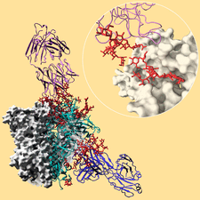
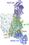
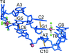
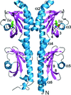
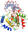
Cover illustration: Broadly neutralizing antibodies PGT128 (purple ribbon) and 8ANC195 (blue ribbon) interact with the glycan shield (red ball-and-stick) of the HIV-1 envelope glycoprotein trimer (Kong et al., p. 2099). Antibody interactions with one protomer of the HIV-1 trimer are shown. Each protomer (turquoise ribbon) is comprised of gp120 and gp41. The other two protomers of the trimer are displayed as white molecular surfaces. The inset zooms in on the interaction between PGT128 (purple ribbon) and gp120 (white molecular surface). PGT128 allosterically interacts with the glycan shield of the HIV-1 envelope glycoprotein trimer. PGT128 contacts an N-linked glycan at position Asn301 on the HIV-1 trimer, which then alters the conformation of the N-linked glycan at position Asn262, which differs from the unliganded Asn262 glycan conformation. The information derived from this crystal structure is being used for design of HIV-1 vaccine immunogens. This illustration was generated by Christina Corbaci and Leopold Kong.

research papers


link to html
A highly X-ray-transparent, silicon nitride-based device has been designed and fabricated to harvest protein microcrystals for high-resolution X-ray diffraction data collection using microfocus beamlines and XFELs.

link to html
The crystal structure of an Rsd–HPr complex was determined in order to reveal the binding interface between Rsd and HPr. A molecular link between global gene regulation and nutrient availability is described.


link to html
Lattice vibrations account for the structured component of the diffuse scatter produced by crystals of the 70S ribosome from T. thermophilus.

link to html
Nicotinamide mononucleotide adenylyltransferase (NMNAT) catalyzes the biosynthesis of NAD+ and NaAD+. Surprisingly, when expressed in E. coli, NMNAT mutants trapped a molecule of NADP+ in their active sites. This NADP+ was bound in a conformation that was quite different from that displayed by NAD+ in the native enzyme complex.

link to html
A combination of crystal-contact engineering and surface-entropy reduction was used to enable the crystallization and subsequent structural solution of two spliceosomal assembly intermediates.


link to html
In vitro neuromuscular studies were performed to demonstrate that suramin neutralizes the toxic activity of a PLA2-like protein, corroborating calorimetric assays. Crystallographic studies demonstrated that the inhibitor binds to the surface of the toxin, obstructing its membrane docking and disruption sites and confirming the proposed myotoxic mechanism.

link to html
A method is reported for collecting room-temperature data from protein crystals under vacuum by protecting them with a thin graphene layer.

link to html
Epsilon-class glutathione S-transferases (GSTs) are found in invertebrates, including many insects, where they play roles in the detoxication of insecticides and in development. The crystal structures of two epsilon-class GSTs from D. melanogaster are reported and compared to aid in the characterization of the functional differences between the isozymes.

link to html
The crystal structure of the broadly neutralizing antibodies 8ANC195 and PGT128 bound to an HIV-1 envelope trimer has been determined. Structural and binding analyses have elucidated the full epitopes for these antibodies in the context of the intact viral glycoprotein, providing improved templates for HIV-1 vaccine design.

link to html
The crystal structures of two constructs of RC1339/APRc from R. conorii, consisting of either residues 105–231 or 110–231 followed by a His tag, have been determined in three different crystal forms. The monomer of APRc assumes a retropepsin-like fold, but the dimer observed in all crystal forms is entirely different from the canonical dimer of retropepsins.

link to html
The self-complementary DNA decamer d(GCATGCATGC) crystallizes as an unusual double-folded structure, which serves as the basis for a possible structural model of triplet-repeat DNA sequences.

link to html
Structural insight is provided into the mechanism by which a widespread family of bacterial chemotaxis receptors containing the periplasmic tandem Per–Arnt–Sim (PAS) sensing domain recognize free amino acids, and a model for signal transduction across the membrane is proposed.

link to html
The high-resolution structure of uracil-DNA N-glycosylase (UNG) from D. radiodurans in complex with DNA provides support for a model in which DNA compression and minor-groove stabilization is crucial for the uracil damage-detection mechanism of UNGs.
Follow Acta Cryst. D
Sign up for e-alerts
Follow Acta Cryst. on Twitter
Follow us on facebook
Sign up for RSS feeds