
Acta Crystallographica
Section E CRYSTALLOGRAPHIC COMMUNICATIONS


 menu


research communications
| CRYSTALLOGRAPHIC COMMUNICATIONS |
 access
Synthesis and structure of poly[hydroxonium [μ4-bromido-tetra-μ3-bromido-diplumbate(II)]]

aDepartment of Chemistry, Taras Shevchenko National University of Kyiv, Volodymyrska st. 64/13, 01601 Kyiv, Ukraine, and bDepartment of Inorganic Polymers, "Petru Poni" Institute of Macromolecular Chemistry, Aleea Grigore Ghica Voda 41A, 700487 Iasi, Romania
*Correspondence e-mail: [email protected]
The title compound, {(H3O)[Pb2Br5]}n, crystallizes in the tetragonal space group I4/mcm. The asymmetric unit comprises one Pb, two Br and one O sites with site symmetry m.2m, 4/m.., ..m and 422, respectively. Bromide ions link the PbII atoms to form a two-periodic Pb2Br5− anion extending parallel to the ab plane, whilst hydroxonium cations are embedded between neighbouring inorganic layers. Although the hydrogen positions of the hydroxonium cation could not be determined, close O⋯Br contacts suggest possible O—H⋯Br hydrogen-bonding interactions between cations and the anionic layers.
Keywords: hydronium bromidoplumbate(II); lead(II) bromide; two-periodic network; tetragonal crystal structure; Pb—Br coordination; disorder.
CCDC reference: 2599077
1. Chemical context
Lead(II) halide and halidoplumbate(II) compounds display various coordination environments and structural connectivities, both of which are influenced by the stereochemical activity of the PbII 6s2 lone pair and by the terminal or bridging character of the halido ligands (Shimoni-Livny et al., 1998 ; Mitzi, 2001). Lead(II) halides are widely used as precursors for hybrid lead-halide perovskites. The latter have attracted considerable interest due to their semiconducting properties and potential applications in photovoltaics and optoelectronics (Yuan et al., 2025; Kucheriv et al., 2023, 2025). The chemistry of halidoplumbate species in solution is also central to the formation of lead-halide perovskites. Studies of lead-halide precursor solutions and solvent-dependent phase formation highlight the importance of halidoplumbate speciation and the coordination environment of PbII (Sharenko et al., 2017). Hydroxonium ions can exhibit different hydrogen-bonding arrangements in crystalline solids, with their local structure influenced by interactions with the surrounding atoms (Bernal, 2006).
In this paper, the crystal structure of the title compound, (H3O)Pb2Br5, (I), containing a two-periodic Pb–Br network, is reported.
2. Structural commentary
The of (I) comprises four sites, Pb1, Br1, Br2 and O1. They occupy the special Wyckoff positiosn 8 h, 4 c, 16 l and 4 a, with site symmetry m.2m, 4/m.., ..m and 422, respectively, in the tetragonal I4/mcm. The asymmetric unit, together with selected symmetry-generated bromide ions that complete the local environment of Pb1, is shown in Fig. 1. The Pb1 atom is eight-coordinated by bromide ions, with Pb1—Br2 and Pb1—Br1 distances of 2.8863 (10)/3.3470 (9) and 3.1287 (2) Å, respectively. The Br—Pb1—Br bond angles range from 66.989 (2)° for Br2( − x,
 + y, 1 − z)—Pb1—Br2( + x, − y, 1 − z) to 146.5601 (14)° for Br2—Pb1—Br2( + x, − y, 1 − z). A continuous shape-measure analysis using SHAPE 2.1 (Llunell et al., 2013) identifies a distorted biaugmented trigonal prism (BTPR-8) as the closest ideal figure for the PbBr8 (CShM = 1.278). Bromide anions link neighbouring PbII atoms into a two-periodic Pb2Br5− anion extending parallel to the ab plane (Figs. 2, 3).
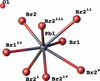 | Figure 1 Fragment of the crystal structure showing the coordination environment of Pb1 in the title compound. Displacement ellipsoids are drawn at the 50% probability level. [Symmetry codes: (i) + y, − + x, 1 − z; (ii) 1 + y, 1 − x, z; (iii) 1 − y, x, z; (iv) − x, + y, 1 − z; (v) + x, − y, 1 − z.] |
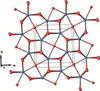 | Figure 2 Fragment of the two-periodic Pb–Br network extending parallel to the ab plane. |
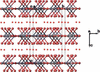 | Figure 3 View of the crystal packing, showing the Pb2Br5− anion layers and the O1-centred hydroxonium ion (small red spheres) between neighbouring layers. |
3. Supramolecular features
The O1-centred hydroxonium ion is embedded between two neighbouring Pb–Br layers and forms fourselected O1⋯Br contacts with bromide ions of symmetry-related Pb—Br units (Fig. 4). These contacts involve both the Br1 and Br2 sites. The observed O1⋯Br contacts (Table 1) suggest possible O—H⋯Br hydrogen-bonding interactions between the hydroxonium ion and the anionic layers. However, the hydrogen atom(s) could not be located from the available X-ray data and were therefore omitted from the refinement model. Consequently, the O—H bond orientations and further numerical details of the hydrogen-bonding scheme remain undetermined, and no specific hydrogen-bonding topology is assigned. A view of the overall crystal packing, including the two-periodic Pb–Br networks and the O1-centred hydroxonium ion, is shown in Fig. 3.
| ||||||||||||||
 ; (ii)
 ; (iii)
 .
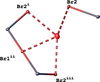 | Figure 4 Local environment of the O1-centred hydroxonium ion, showing four selected O1⋯Br contacts with bromide ions of neighbouring Pb–Br layers. Dashed lines indicate O1⋯Br contacts associated with possible O—H⋯Br hydrogen-bonding; hydrogen atom positions remain undetermined. [Symmetry codes: (i) 1 − x, y, − z; (ii) x − , y + , z - 1/2; (iii) x, 1 − y, − z.] |
4. Database survey
A search of the Crystallography Open Database (COD; Gražulis et al., 2012) identified related PbII bromide compounds, of which two were selected for structural comparison: the hydrated lead(II) bromide phase 3PbBr2·2H2O (Liu et al., 2018) and the oxyhydroxybromide Pb2OOHBr (Kunisawa et al., 2026). The former contains eight-coordinate PbII atoms with PbBr8 or PbBr7O coordination environments, whereas the latter contains PbBr4O4 coordination polyhedra within a Pb–O–Br framework.
5. Synthesis and crystallization
Lead(II) bromide (50 mg, 0.136 mmol) was dissolved in concentrated hydrobromic acid (0.5 ml). After 2 days, light-yellow single crystals suitable for single-crystal X-ray diffraction were formed. They were stored in the mother liquor prior to measurement.
6. Refinement
Crystal data, data collection and structure details are summarized in Table 2. Due to the site symmetry (422) of the oxygen atom, the hydrogen-atom positions of the hydroxonium cation are orientationally disordered. This fact, combined with the high absorption coefficient of (I), has made it impossible to determine the relevant H positions from difference-Fourier syntheses. Therefore, they were not included in the final model; the contribution from the H atoms is accounted for in the molecular weight given in Table 2.
|
Supporting information
CCDC reference: 2599077
contains datablock I. DOI: https://doi.org/10.1107/S2056989026011291/wm5818sup1.cif
Structure factors: contains datablock I. DOI: https://doi.org/10.1107/S2056989026011291/wm5818Isup2.hkl
| (H3O)[Pb2Br5] | Dx = 5.475 Mg m−3 |
| Mr = 832.95 | Mo Kα radiation, λ = 0.71073 Å |
| Tetragonal, I4/mcm | Cell parameters from 1179 reflections |
| a = 8.3948 (4) Å | θ = 2.8–29.7° |
| c = 14.3392 (10) Å | µ = 52.98 mm−1 |
| V = 1010.52 (12) Å3 | T = 100 K |
| Z = 4 | Plate, light yellow |
| F(000) = 1400 | 0.20 × 0.17 × 0.06 mm |
| XtaLAB Synergy, Dualflex, HyPix diffractometer | 368 independent reflections |
| Radiation source: micro-focus sealed X-ray tube, PhotonJet (Mo) X-ray Source | 317 reflections with I > 2σ(I) |
| Mirror monochromator | Rint = 0.048 |
| Detector resolution: 10.0000 pixels mm-1 | θmax = 29.8°, θmin = 2.8° |
| ω scans | h = −11→11 |
| Absorption correction: analytical [CrysAlisPro (Rigaku OD, 2024), based on expressions derived by Clark & Reid, 1995] | k = −10→7 |
| Tmin = 0.011, Tmax = 0.094 | l = −18→13 |
| 1765 measured reflections |
| Refinement on F2 | H-atom parameters not defined |
| Least-squares matrix: full | w = 1/[σ2(Fo2) + (0.037P)2 + 12.0445P] where P = (Fo2 + 2Fc2)/3 |
| R[F2 > 2σ(F2)] = 0.028 | (Δ/σ)max = 0.001 |
| wR(F2) = 0.075 | Δρmax = 2.31 e Å−3 |
| S = 1.09 | Δρmin = −1.59 e Å−3 |
| 368 reflections | Extinction correction: SHELXL2018/3 (Sheldrick 2015b), Fc*=kFc[1+0.001xFc2λ3/sin(2θ)]-1/4 |
| 16 parameters | Extinction coefficient: 0.00046 (9) |
| 0 restraints |
Geometry. All esds (except the esd in the dihedral angle between two l.s. planes) are estimated using the full covariance matrix. The cell esds are taken into account individually in the estimation of esds in distances, angles and torsion angles; correlations between esds in cell parameters are only used when they are defined by crystal symmetry. An approximate (isotropic) treatment of cell esds is used for estimating esds involving l.s. planes. |
| x | y | z | Uiso*/Ueq | ||
| Pb1 | 0.83338 (4) | 0.33338 (4) | 0.500000 | 0.0116 (2) | |
| Br1 | 1.000000 | 0.000000 | 0.500000 | 0.0102 (4) | |
| Br2 | 0.65560 (8) | 0.15560 (8) | 0.36269 (7) | 0.0109 (3) | |
| O1 | 0.500000 | 0.500000 | 0.250000 | 0.030 (4) |
| U11 | U22 | U33 | U12 | U13 | U23 | |
| Pb1 | 0.0094 (3) | 0.0094 (3) | 0.0161 (4) | −0.0005 (2) | 0.000 | 0.000 |
| Br1 | 0.0078 (5) | 0.0078 (5) | 0.0150 (9) | 0.000 | 0.000 | 0.000 |
| Br2 | 0.0105 (4) | 0.0105 (4) | 0.0117 (5) | −0.0014 (4) | −0.0010 (3) | −0.0010 (3) |
| O1 | 0.034 (6) | 0.034 (6) | 0.023 (8) | 0.000 | 0.000 | 0.000 |
| Pb1—Br1 | 3.1287 (2) | Pb1—Br2ii | 2.8863 (10) |
| Pb1—Br1i | 3.1287 (2) | Pb1—Br2 | 2.8863 (10) |
| O1···Br1iii | 3.5848 (3) | O1···Br2iv | 3.5604 (1) |
| O1···Br2 | 3.5604 (1) | O1···Br2v | 3.5604 (1) |
| Br1—Pb1—Br1i | 143.113 (17) | Pb1—Br1—Pb1vi | 180.0 |
| Br2ii—Pb1—Br1i | 76.624 (9) | Pb1vii—Br1—Pb1viii | 180.0 |
| Br2ii—Pb1—Br1 | 76.624 (9) | Pb1—Br1—Pb1vii | 90.0 |
| Br2—Pb1—Br1 | 76.624 (9) | Pb1vii—Br1—Pb1vi | 90.0 |
| Br2—Pb1—Br1i | 76.624 (9) | Pb1viii—Br1—Pb1vi | 90.0 |
| Br2ii—Pb1—Br2 | 86.02 (4) | Pb1—Br1—Pb1viii | 90.0 |
| Symmetry codes: (i) −x+3/2, y+1/2, −z+1; (ii) x, y, −z+1; (iii) x−1/2, y+1/2, z−1/2; (iv) −x+1, y, −z+1/2; (v) x, −y+1, −z+1/2; (vi) −x+2, −y, −z+1; (vii) y+1, −x+1, −z+1; (viii) −y+1, x−1, z. |
Acknowledgements
The authors are grateful to the FAIRE programme provided by the Cambridge Crystallographic Data Centre (CCDC) for the opportunity to use the Cambridge Structural Database (CSD) and associated software.
Funding information
Funding for this research was provided by: Ministry of Education and Science of Ukraine (grant Nos. 24BF037-01M and 24BF037-02).
References
 Bernal, I. (2006). C. R. Chim. 9, 1454–1466. Web of Science CrossRef CAS Google Scholar
Clark, R. C. & Reid, J. S. (1995). Acta Cryst. A51, 887–897. CrossRef CAS Web of Science IUCr Journals Google Scholar
Dolomanov, O. V., Bourhis, L. J., Gildea, R. J., Howard, J. A. K. & Puschmann, H. (2009). J. Appl. Cryst. 42, 339–341. Web of Science CrossRef CAS IUCr Journals Google Scholar
Gražulis, S., Daškevič, A., Merkys, A., Chateigner, D., Lutterotti, L., Quirós, M., Serebryanaya, N. R., Moeck, P., Downs, R. T. & Le Bail, A. (2012). Nucleic Acids Res. 40, D420–D427. Web of Science PubMed Google Scholar
Kucheriv, O. I., Haleliuk, D. A., Shova, S. & Gural'skiy, I. A. (2025). Dalton Trans. 54, 1618–1624. CSD CrossRef CAS PubMed Google Scholar
Kucheriv, O. I., Sirenko, V. Y., Petrosova, H. R., Pavlenko, V. A., Shova, S. & Gural'skiy, I. A. (2023). Inorg. Chem. Front. 10, 6953–6963. Web of Science CSD CrossRef CAS Google Scholar
Kunisawa, H., Yamaura, J.-I. & Nomura, T. (2026). IUCrData 11, x260790. Google Scholar
Liu, M., Zhao, J., Luo, Z., Sun, Z., Pan, N., Ding, H. & Wang, X. (2018). Chem. Mater. 30, 5846–5852. CSD CrossRef ICSD CAS Google Scholar
Llunell, M., Casanova, D., Cirera, J., Alemany, P. & Alvarez, S. (2013). SHAPE. Version 2.1. Universitat de Barcelona, Barcelona, Spain. Google Scholar
Mitzi, D. B. (2001). J. Chem. Soc. Dalton Trans. pp. 1–12. Web of Science CrossRef Google Scholar
Rigaku OD (2024). CrysAlis PRO. Rigaku Oxford Diffraction, Yarnton, England. Google Scholar
Sharenko, A., Mackeen, C., Jewell, L., Bridges, F. & Toney, M. F. (2017). Chem. Mater. 29, 1315–1320. CrossRef CAS Google Scholar
Sheldrick, G. M. (2015a). Acta Cryst. A71, 3–8. Web of Science CrossRef IUCr Journals Google Scholar
Sheldrick, G. M. (2015b). Acta Cryst. C71, 3–8. Web of Science CrossRef IUCr Journals Google Scholar
Shimoni-Livny, L., Glusker, J. P. & Bock, C. W. (1998). Inorg. Chem. 37, 1853–1867. Web of Science CrossRef CAS Google Scholar
Westrip, S. P. (2010). J. Appl. Cryst. 43, 920–925. Web of Science CrossRef CAS IUCr Journals Google Scholar
Yuan, L., Xue, Q., Wang, F., Li, N., Waterhouse, G. I. N., Brabec, C. J., Gao, F. & Yan, K. (2025). Chem. Rev. 125, 5057–5162. CrossRef CAS PubMed Google Scholar
This is an open-access article distributed under the terms of the Creative Commons Attribution (CC-BY) Licence, which permits unrestricted use, distribution, and reproduction in any medium, provided the original authors and source are cited.
| CRYSTALLOGRAPHIC COMMUNICATIONS |