
Acta Crystallographica Section F
Acta Crystallographica
Section F STRUCTURAL BIOLOGY COMMUNICATIONS
Acta Crystallographica
Section F STRUCTURAL BIOLOGY COMMUNICATIONS


 menu
|
|
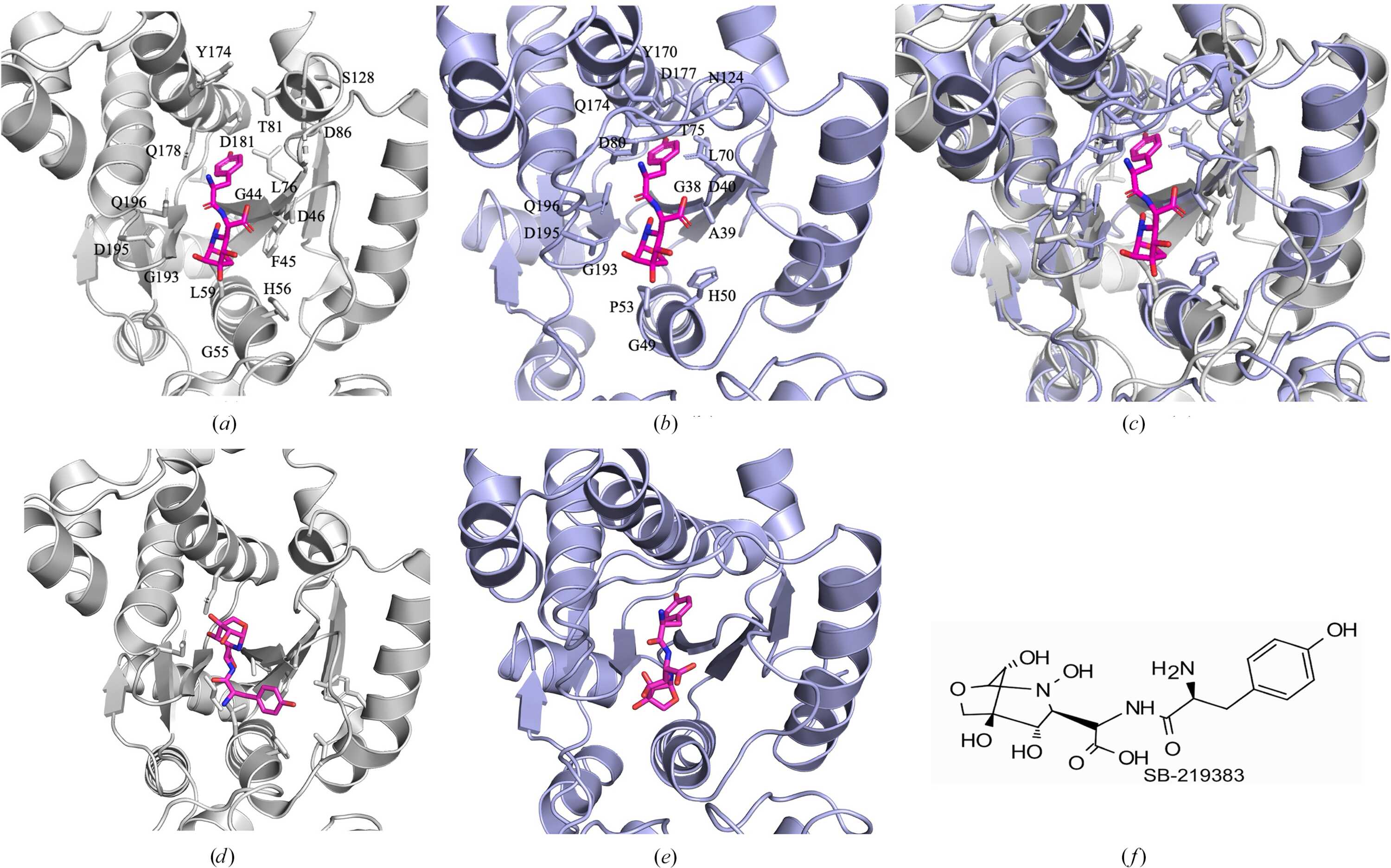
Figure 2
Superposed and modelled SB-219383 inhibitor binding. (a) HpTyrRS has conserved amino-acid residues in the superposed SB-219383 binding cavity. (b) SB-219383 (magenta) binds to SaTyrRS (blue) residues shown as blue sticks, as identified from PDB entry 1jii. (c) Superposed HpTyrRS (gray) and SaTyrRS (blue) reveal conserved amino acids. (d) Top docking pose of SB-219383 with HpTyrRS; docking score −8.03. (e) Top docking pose of SB-219383 with SaTyrRS; docking score −8.28. (f) CHEMDRAW representation of SB-219383. HpTyrRS (PDB entry 6byq) was superposed with SaTyrRS (PDB entry 1jii) within PyMOL to generate (a)–(c). (a)–(e) are in the same orientation as Fig. 1  and are magnified to visualize SB-219383. All figures were generated with PyMOL unless otherwise stated. |
| STRUCTURAL BIOLOGY COMMUNICATIONS |
ISSN: 2053-230X
Open  access