
Acta Crystallographica
Section F STRUCTURAL BIOLOGY COMMUNICATIONS


 menu


research communications
| STRUCTURAL BIOLOGY COMMUNICATIONS |
 access
of tyrosine-tRNA synthetase from Helicobacter pylori G27

aDartmouth College, Hanover, New Hampshire, USA, bUniversity of California, Berkeley, Berkeley, California, USA, cCenter for Global Infectious Disease Research, Seattle Children's Research Institute, 307 Westlake Avenue North, Suite 500, Seattle, WA 98109, USA, dSeattle Structural Genomics Center for Infectious Diseases, Seattle, Washington, USA, eUCB BioSciences, Bainbridge Island, Washington, USA, fChemistry and Biochemistry, Hampton University, Hampton, VA 23666, USA, gDartmouth Cancer Center, Lebanon, New Hampshire, USA, and hBiochemistry and Cell Biology, Dartmouth Geisel School of Medicine, New Hampshire, USA
*Correspondence e-mail: [email protected], [email protected]
This article is part of a special issue celebrating early career researchers in structural science.
Helicobacter pylori, a major cause of gastric ulcers and cancers, infects nearly half of the world's population. The crystal structure of a potential H. pylori structure-guided drug-discovery target, tyrosine-tRNA synthetase (HpTyrRS), has been determined by the Seattle Structural Genomics Center for Infectious Disease (SSGCID). HpTyrRS is an enzyme that catalyzes the ATP-dependent attachment of tyrosine to tRNA. HpTyrRS was produced and crystallized, and its structure was determined at 2.2 Å resolution. HpTyrRS has a prototypical bacterial TyrRS topology and forms dimers in solution. HpTyrRS shares a conserved binding cavity for a previously identified Staphylococcus aureus TyrRS inhibitor (SB-219383). The reported structure and analysis establish HpTyrRS as a potential target for structure-guided drug discovery and repurposing.
PDB reference: tyrosine-tRNA synthetase from Helicobacter pylori G27, 6byq
1. Introduction
Helicobacter pylori is a helical, Gram-negative, microaerophilic bacterium that colonizes the stomach of ∼50% of the world's human population (Costa et al., 2024 ). H. pylori causes chronic gastritis, peptic ulcer disease (PUD), gastric mucosa-associated lymphoid tissue lymphoma (MALT) and gastric adenocarcinoma (Sabbagh et al., 2019). The helical shape of H. pylori facilitates movement in the human body, while the coccoid form allows colonization of the gastric epithelial cells (Al-Fakhrany & Elekhnawy, 2023). H. pylori colonization induces pathological changes in the gastric microenvironment (Wang et al., 2015). Increasing antibiotic resistance of infectious bacteria has been a global concern for decades, prompting the adoption of combination therapy involving two or more types of antibiotics, such as amoxicillin, clarithromycin, metronidazole, tetracycline, levofloxacin and rifabutin, as well as bismuth, to enhance healing and protect the gastric mucosa (Wang et al., 2015). The increasing drug resistance of H. pylori reveals an unmet need for new therapeutics. The Seattle Structural Genomics Center for Infectious Disease (SSGCID) has identified H. pylori aminoacyl-tRNA synthetases, including tyrosine-tRNA synthetase (HpTyrRS), as targets for structure-guided drug discovery and repurposing. TyrRS catalyzes the bonding of tyrosine to tRNA (Xin et al., 2000). Aminoacyl-tRNA synthetases (aaRSs) are enzymes that catalyze the esterification of amino acids to the corresponding tRNA, where the product of the process plays a crucial role in protein synthesis (Rubio Gomez & Ibba, 2020). The aaRS family, specifically TyrRS, have been identified as promising targets for new antibacterials (Zhu et al., 2015; Sun et al., 2017; Monobe et al., 2024). Here, we present the purification, crystallization and structure of HpTyrRS as a first step towards structure-guided inhibitor design.
2. Materials and methods
2.1. Macromolecule production
HpTyrRS was cloned, expressed and purified using previously described methods (Stacy et al., 2011; Serbzhinskiy et al., 2015; Rodríguez-Hernández et al., 2023). The full-length gene for tyrosine-tRNA synthetase (EC 6.1.1.1) from H. pylori G27 (UniProt B5Z7D7) encoding amino acids 1–402 was PCR-amplified from genomic DNA using the primers in Table 1. The gene was cloned into the expression vector BG1861 to generate N-terminally His-tagged plasmid DNA. The plasmid was transformed into chemically competent Escherichia coli BL21(DE3) Rosetta cells. These cells are an institutional variation of the classic Rosetta expression strain that layer a protective phage-resistance mutation onto the rare-codon translation power of the Rosetta strain.
| ||||||||||||||||||
After small-scale expression testing, a 2 l culture was grown in a LEX Bioreactor (Epiphyte Three) using auto-induction media (Studier, 2005) and standardized SSGCID methods (Serbzhinskiy et al., 2015). HpTyrRS was purified using the previously described two-step protocol of an immobilized metal (Ni2+)-affinity chromatography (IMAC) step followed by size-exclusion chromatography (SEC) on an ÄKTApurifier 10 (GE Healthcare) using automated IMAC and SEC programs (Serbzhinskiy et al., 2015). Briefly, thawed bacterial pellets (25 g) were lysed by sonication in 200 ml lysis buffer [20 mM HEPES pH 7.0, 300 mM NaCl, 5%(v/v) glycerol, 0.5%(w/v) CHAPS, 30 mM imidazole, 10 mM MgCl2 and five tablets of protease inhibitor cocktail (cOmplete Mini, EDTA-free Roche, Basel, Switzerland)]. After sonication, the crude lysate was incubated with 20 µl (25 units per µl) benzonase while mixing for 45 min at room temperature. The lysate was centrifuged at 5000g for 1 h to remove insoluble debris. The clarified supernatant was passed over an Ni–NTA HisTrap FF 5 ml column (GE Healthcare), which had been pre-equilibrated with wash buffer [20 mM HEPES pH 7.0, 300 mM NaCl, 5%(v/v) glycerol, 30 mM imidazole]. The column was washed with 20 column volumes (CV) of wash buffer and eluted with elution buffer [20 mM HEPES pH 7.0, 300 mM NaCl, 5%(v/v) glycerol, 500 mM imidazole] over a 7 CV linear gradient. Peak fractions were pooled and concentrated to a final volume of 5 ml for SEC. The 5 ml protein sample was loaded onto a Superdex 75 26/60 column (GE Biosciences) attached to an ÄKTAprime plus FPLC system (GE Biosciences) that had been pre-equilibrated with SEC buffer (20 mM HEPES pH 7.0, 300 mM NaCl, 5% glycerol, 1 mM TCEP). HpTyrRS eluted as a single, symmetrical, monodisperse peak. The peak fractions were collected and assessed for purity by SDS–PAGE on a 4–20% protein gel (Invitrogen) and visualized by staining with InstantBlue colloidal stain (Expedeon, San Diego, California. USA). Recombinant HpTyrRS was ∼95% pure. The peak fraction was pooled and concentrated to ∼20 mg ml−1 using an Amicon purification system (Millipore). Aliquots were flash-frozen in liquid nitrogen and stored at −80°C until use.
2.2. Crystallization
HpTyrRS was screened for crystallization conditions using the Molecular Dimensions Morpheus commercial screen. Crystals were grown in Molecular Dimensions Morpheus condition D11 (Table 2) and did not require additional cryoprotectant prior to data collection.
| ||||||||||||||||||||
2.3. Data collection and processing
Data were collected as reported in Table 3. Data were integrated with XDS and reduced with XSCALE (Kabsch, 2010). Raw X-ray diffraction images are stored at the Integrated Resource for Reproducibility in Macromolecular Crystallography at https://www.proteindiffraction.org.
| ||||||||||||||||||||||||||||||||||||||||||||||
2.4. Structure solution and refinement
The structure of HpTyrRS was determined by molecular replacement with SIMBAD (Simpkin et al., 2018) from the CCP4 suite of programs (Collaborative Computational Project, 1994; Krissinel & Henrick, 2004; Winn et al., 2011;Agirre et al., 2023) using PDB entry 1h3f (Yaremchuk et al., 2002) as the search model. The structure was refined using iterative cycles of phenix.refine (Afonine et al., 2012) followed by manual rebuilding of the structure using Coot (Emsley & Cowtan, 2004; Emsley et al., 2010).
One molecule is present in the asymmetric unit, consistent with a Matthews coefficient of 2.74 Å3 Da−1, corresponding to a 55% solvent content in the protein crystal. The final model included two N-terminal histidine residues from the His-tag and 311 main-chain residues (Fig. 1, Table 4). Six vector-derived residues and 96 main-chain residues were not modeled because they lacked unambiguous electron density at a 0.9 r.m.s.d. cutoff in the 2Fo − Fc electron density after iterative cycles of model building and The structure quality was checked using MolProbity (Williams et al., 2018). Refinement statistics are shown in Table 4. Coordinates and structure factors have been deposited with the Worldwide PDB (wwPDB) as entry pdb_00006byq.
| |||||||||||||||||||||||||||||||||||||||||||||||||||
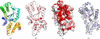 | Figure 1 HpTyrRS structure. (a) HpTyrRS has a prototypical TyrRS topology (colored from blue at the N-terminus to red at the C-terminus). (b) ENDscript ribbon diagram. Ribbon thickness reflects secondary-structure similarity: thinner ribbons indicate more conserved regions and thicker ribbons suggest lower conservation, as determined by r.m.s.d. alignment of structures. The ribbon is colored by sequence identity (red = identical residues). (c) Solvent-accessible surface area of HpTyrRS colored by ENDscript sequence conservation, with identical residues colored red, reveals a large conserved central cavity. (d) SB-219383 in magenta sticks sits in the open central cavity in both SaTyrRS (blue) and HpTyrRS (gray). HpTyrRS (PDB entry 6byq) was aligned with SaTyrRS (PDB entry 1jii) within PyMOL to generate (d). (a)–(d) are in the same orientation. All figures were generated with PyMOL. |
2.5. Inhibitor docking
Inhibitor docking was performed using AutoDock Vina v1.2.7 to determine whether HpTyrRS is suitable for drug repurposing (Eberhardt et al., 2021; Cosconati et al., 2010; Trott & Olson, 2010). UCSF Chimera DockPrep was used to prepare the ligand, add hydrogens and charges, remove solvent molecules and repair incomplete side chains in HpTyrRS (Lang et al., 2009; Moustakas et al., 2006). The docking search space was defined based on residues within 5 Å of SB-219383 in the predicted active site, after structural alignment with Staphylococcus aureus TyrRS (SaTyrRS; PDB entry 1jii; Qiu et al., 2001) within PyMOL. The grid box was centered at X = −17.1375 Å, Y = −20.2976 Å, Z = 14.9006 Å, with box dimensions of 17.6119 × 23.3816 × 26.8822 Å and a grid spacing of 0.375 Å. Docking simulations were conducted with an exhaustiveness of 12, using 8 CPU cores and a verbosity level of 1. As a control, the docking search was repeated with SaTyrRS.
3. Results and discussion
Preparative SEC revealed that HpTyrRS assembles as a ∼77 kDa dimer, consistent with PISA analysis (https://www.ebi.ac.uk/pdbe/pisa/). This dimer suggests a monomer of ∼38 kDa, which is smaller than the expressed protein of 45 kDa. Approximately 24% of the sequence was missing and could not be modeled, notably 83 residues at the carboxyl terminus from Gln319 through Asn402 (Table 1). Additionally, six vector-derived residues −8MAHHHH−3 and seven residues 86DPTGKNE92 could not be modeled. The expected molecular weight of the modeled protein is ∼37 kDa, consistent with SEC data. A possible hypothesis is C-terminal proteolysis. Mass-spectroscopic analysis of recombinant HpTyrRS and HpTyrRS crystals is planned to confirm their amino-acid sequence and predicted molecular mass.
HpTyrRS is a prototypical bacterial TyrRS with the canonical bacterial tyrosine-tRNA (TyrRS) architecture consisting of a compact, globular α/β fold comprising 19 α-helices and six β-strands, organized into three βαβ units, one β-bulge and a single β-sheet (Fig. 1). ENDscript analysis (Gouet et al., 1999, 2003) revealed that HpTyrRS shares extensive secondary-structural similarity with other bacterial TyrRS, and the closest structural homologs of HpTyrRS are TyrRS from Acinetobacter baumannii (PDB entry 6bqy), Thermus thermophilus (PDB entry 1h3e) and E. coli (PDB entry 1vbm) (Yaremchuk et al., 2002; Kobayashi et al., 2005; Figs. 1b, 1c and Supplementary Fig. S1). Similarly, PDBeFold analysis (https://www.ebi.ac.uk/msd-srv/ssm; Krissinel & Henrick, 2004) with a default threshold cutoff of 70% identified the closest structural neighbor of HpTyrRS as the A. baumannii TyrRS structure (PDB entries 6bqy and 6bqz), with ∼57% sequence identity and ∼1.6 Å r.m.s.d for alignment of all main-chain atoms. Likewise, a DALI search for similar structures against the PDB was consistent with PDBeFold and established HpTyrRS as a prototypical bacterial TyrRS, supporting the feasibility of inhibitor repurposing from other bacterial TyrRS.
To assess the feasibility of future drug repurposing of HpTyrRS, (2S)-2-[[(2S)-2-amino-3-(4-hydroxyphenyl)propanoyl]amino]-2-[(1S,3S,4S,5R,8R)-2,4,5,8-tetrahydroxy-7-oxa-2-azabicyclo[3.2.1]octan-3-yl]acetic acid; SB-219383; Fig. 2d), a competitive, selective and potent inhibitor of bacterial TyrRS, was docked to HpTyrRS. As a control, SB-219383 was docked back into SaTyrRS. SB-219383 is a potent (IC50 of ∼0.5 nM) SaTyrRS inhibitor produced by Micromonospora, and a weaker inhibitor of mammalian TyrRS, specifically with an IC50 of ∼2 µM for the rat liver orthologue (Stefanska et al., 2000; Greenwood & Gentry, 2002). This selectivity profile strengthens the rationale for testing SB-219383 against HpTyrRS.
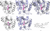 | Figure 2 Superposed and modelled SB-219383 inhibitor binding. (a) HpTyrRS has conserved amino-acid residues in the superposed SB-219383 binding cavity. (b) SB-219383 (magenta) binds to SaTyrRS (blue) residues shown as blue sticks, as identified from PDB entry 1jii. (c) Superposed HpTyrRS (gray) and SaTyrRS (blue) reveal conserved amino acids. (d) Top docking pose of SB-219383 with HpTyrRS; docking score −8.03. (e) Top docking pose of SB-219383 with SaTyrRS; docking score −8.28. (f) CHEMDRAW representation of SB-219383. HpTyrRS (PDB entry 6byq) was superposed with SaTyrRS (PDB entry 1jii) within PyMOL to generate (a)–(c). (a)–(e) are in the same orientation as Fig. 1 and are magnified to visualize SB-219383. All figures were generated with PyMOL unless otherwise stated. |
The structure of SaTyrRS with bound SB-219383 has been reported as PDB entry 1jii (Qiu et al., 2001). SaTyrRS shares ∼26% sequence identity with HpTyrRS and modest structural alignment (r.m.s.d. 2.9 Å, alignment length 295 residues; Fig. 1d). SB-219383 sits in a central binding cavity in SaTyrRS (Figs. 1d, 2b and 2c). The central binding cavity of SaTyrRS shares similar amino acids with HpTyrRS (Figs. 2a–2c). Many residues within 5 Å of SB-219383 are conserved (Fig. 3 and Table 5). Additionally, some of these residues are conserved in bacterial TyrRS (Supplementary Fig. S1). The top docking pose of SB-219383 into HpTyrRS (Fig. 2d) does not resemble the superposed orientation (Fig. 2a). The control docking of SB-219383 into SaTyrRS (Fig. 2e) more closely resembles the crystal structure (Fig. 2b) than the top pose obtained from docking into HpTyrRS (Fig. 2d), which may reflect the cavity's large open size and suggest the need for more experimental data to define inhibitor binding by HpTyrRS.
| ||||||||||||||||||||||||||||||||||||||||||||
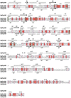 | Figure 3 Structure-based primary-sequence alignment of HpTyrRS (PDB entry 6byq) and SaTyrRS (PDB entry 1jii). The secondary-structure elements are as follows: α-helices are shown as large coils, 310-helices as small coils labeled η and β-strands as arrows labeled β, with β-turns labeled TT. Identical residues are shown in white font on a red background, while conserved residues are shown in red font and conserved regions are shown in blue boxes. The residues interacting with SB-219383 are indicated with green boxes. This figure was generated using ESPript 3.0 with alignment in Clustal Omega. |
4. Conclusion
HpTyrRS is a prototypical bacterial TyrRS with a large central pocket. Computational docking suggests that the pocket may be capable of binding validated SaTyrRS inhibitors such as SB-219383 and may facilitate structure-guided H. pylori drug discovery and repurposing. Future studies will determine the activity of HpTyrRS and assess inhibition by known bacterial TyrRS inhibitors including SB-219383.
Supporting information
Link https://proteindiffraction.org/project/6byq/
Raw X-ray diffraction images
Supplementary Fig S1. DOI: https://doi.org/10.1107/S2053230X26010253/rf5052sup1.pdf
Acknowledgements
David Igbalajobi is a 2026 Program for Oncology Workforce Education and Research Experience at Dartmouth (POWERED) scholar funded by Friends of the Dartmouth Cancer Center. Jesuferanmi Ayanlade is a 2024 POWERED scholar. Jason Taylor is a Dartmouth 2025–2027 American Cancer Society Individualized Mentorship Program to Accelerate Cancer Training (IMPACT) post-baccalaureate fellow. Raymond Gao is a 2025 American Chemical Society Project SEED Fellow.
Funding information
Funding for this research was provided by: National Institute of Allergy and Infectious Diseases (contract No. 75N93022C00036 to Peter J. Myler, Bart Staker); American Cancer Society (grant No. 1310983 to Oluwatoyin A. Asojo).
References
 Afonine, P. V., Grosse-Kunstleve, R. W., Echols, N., Headd, J. J., Moriarty, N. W., Mustyakimov, M., Terwilliger, T. C., Urzhumtsev, A., Zwart, P. H. & Adams, P. D. (2012). Acta Cryst. D68, 352–367. Web of Science CrossRef CAS IUCr Journals Google Scholar
Agirre, J., Atanasova, M., Bagdonas, H., Ballard, C. B., Baslé, A., Beilsten-Edmands, J., Borges, R. J., Brown, D. G., Burgos-Mármol, J. J., Berrisford, J. M., Bond, P. S., Caballero, I., Catapano, L., Chojnowski, G., Cook, A. G., Cowtan, K. D., Croll, T. I., Debreczeni, J. É., Devenish, N. E., Dodson, E. J., Drevon, T. R., Emsley, P., Evans, G., Evans, P. R., Fando, M., Foadi, J., Fuentes-Montero, L., Garman, E. F., Gerstel, M., Gildea, R. J., Hatti, K., Hekkelman, M. L., Heuser, P., Hoh, S. W., Hough, M. A., Jenkins, H. T., Jiménez, E., Joosten, R. P., Keegan, R. M., Keep, N., Krissinel, E. B., Kolenko, P., Kovalevskiy, O., Lamzin, V. S., Lawson, D. M., Lebedev, A. A., Leslie, A. G. W., Lohkamp, B., Long, F., Malý, M., McCoy, A. J., McNicholas, S. J., Medina, A., Millán, C., Murray, J. W., Murshudov, G. N., Nicholls, R. A., Noble, M. E. M., Oeffner, R., Pannu, N. S., Parkhurst, J. M., Pearce, N., Pereira, J., Perrakis, A., Powell, H. R., Read, R. J., Rigden, D. J., Rochira, W., Sammito, M., Sánchez Rodríguez, F., Sheldrick, G. M., Shelley, K. L., Simkovic, F., Simpkin, A. J., Skubak, P., Sobolev, E., Steiner, R. A., Stevenson, K., Tews, I., Thomas, J. M. H., Thorn, A., Valls, J. T., Uski, V., Usón, I., Vagin, A., Velankar, S., Vollmar, M., Walden, H., Waterman, D., Wilson, K. S., Winn, M. D., Winter, G., Wojdyr, M. & Yamashita, K. (2023). Acta Cryst. D79, 449–461. Web of Science CrossRef IUCr Journals Google Scholar
Al-Fakhrany, O. M. & Elekhnawy, E. (2023). Arch. Microbiol. 205, 301. PubMed Google Scholar
Chen, V. B., Arendall, W. B., Headd, J. J., Keedy, D. A., Immormino, R. M., Kapral, G. J., Murray, L. W., Richardson, J. S. & Richardson, D. C. (2010). Acta Cryst. D66, 12–21. Web of Science CrossRef CAS IUCr Journals Google Scholar
Collaborative Computational Project, Number 4 (1994). Acta Cryst. D50, 760–763. CrossRef Web of Science IUCr Journals Google Scholar
Cosconati, S., Forli, S., Perryman, A. L., Harris, R., Goodsell, D. S. & Olson, A. J. (2010). Exp. Opin. Drug. Discov. 5, 597–607. CrossRef Google Scholar
Costa, L., das Graças Carvalho, M., La Guárdia Custódio Pereira, A. C., Teixeira Neto, R. G., Andrade Figueiredo, L. C. & Barros-Pinheiro, M. (2024). Med. Princ. Pract. 33, 173–184. CrossRef PubMed Google Scholar
Eberhardt, J., Santos-Martins, D., Tillack, A. F. & Forli, S. (2021). J. Chem. Inf. Model. 61, 3891–3898. Web of Science CrossRef CAS PubMed Google Scholar
Emsley, P. & Cowtan, K. (2004). Acta Cryst. D60, 2126–2132. Web of Science CrossRef CAS IUCr Journals Google Scholar
Emsley, P., Lohkamp, B., Scott, W. G. & Cowtan, K. (2010). Acta Cryst. D66, 486–501. Web of Science CrossRef CAS IUCr Journals Google Scholar
Engh, R. A. & Huber, R. (2006). International Tables for Crystallography, Vol. F, 1st online ed., edited by M. G. Rossmann & E. Arnold, pp. 382–392. Chester: International Union of Crystallography. Google Scholar
Gouet, P., Courcelle, E., Stuart, D. I. & Métoz, F. (1999). Bioinformatics, 15, 305–308. Web of Science CrossRef PubMed CAS Google Scholar
Gouet, P., Robert, X. & Courcelle, E. (2003). Nucleic Acids Res. 31, 3320–3323. Web of Science CrossRef PubMed CAS Google Scholar
Greenwood, R. C. & Gentry, D. R. (2002). J. Antibiot. 55, 423–426. CrossRef Google Scholar
Kabsch, W. (2010). Acta Cryst. D66, 125–132. Web of Science CrossRef IUCr Journals Google Scholar
Kobayashi, T., Takimura, T., Sekine, R., Kelly, V. P., Kamata, K., Sakamoto, K., Nishimura, S. & Yokoyama, S. (2005). J. Mol. Biol. 346, 105–117. CrossRef PubMed CAS Google Scholar
Krissinel, E. & Henrick, K. (2004). Acta Cryst. D60, 2256–2268. Web of Science CrossRef CAS IUCr Journals Google Scholar
Lang, P. T., Brozell, S. R., Mukherjee, S., Pettersen, E. F., Meng, E. C., Thomas, V., Rizzo, R. C., Case, D. A., James, T. L. & Kuntz, I. D. (2009). RNA, 15, 1219–1230. Web of Science CrossRef PubMed CAS Google Scholar
Monobe, K., Taniguchi, H. & Aoki, S. (2024). Curr. Comput. Aided Drug. Des. 20, 452–462. CrossRef CAS PubMed Google Scholar
Moustakas, D. T., Lang, P. T., Pegg, S., Pettersen, E., Kuntz, I. D., Brooijmans, N. & Rizzo, R. C. (2006). J. Comput. Aided Mol. Des. 20, 601–619. CrossRef PubMed CAS Google Scholar
Parkinson, G., Vojtechovsky, J., Clowney, L., Brünger, A. T. & Berman, H. M. (1996). Acta Cryst. D52, 57–64. CrossRef CAS Web of Science IUCr Journals Google Scholar
Qiu, X., Janson, C. A., Smith, W. W., Green, S. M., McDevitt, P., Johanson, K., Carter, P., Hibbs, M., Lewis, C., Chalker, A., Fosberry, A., Lalonde, J., Berge, J., Brown, P., Houge–Frydrych, C. S. V. & Jarvest, R. L. (2001). Protein Sci. 10, 2008–2016. Web of Science CrossRef PubMed CAS Google Scholar
Rodríguez-Hernández, D., Vijayan, K., Zigweid, R., Fenwick, M. K., Sankaran, B., Roobsoong, W., Sattabongkot, J., Glennon, E. K. K., Myler, P. J., Sunnerhagen, P., Staker, B. L., Kaushansky, A. & Grøtli, M. (2023). Nat. Commun. 14, 5408. Web of Science PubMed Google Scholar
Rubio Gomez, M. A. & Ibba, M. (2020). RNA, 26, 910–936. Web of Science CrossRef PubMed Google Scholar
Sabbagh, P., Javanian, M., Koppolu, V., Vasigala, V. R. & Ebrahimpour, S. (2019). Eur. J. Clin. Microbiol. Infect. Dis. 38, 1035–1045. CrossRef CAS PubMed Google Scholar
Serbzhinskiy, D. A., Clifton, M. C., Sankaran, B., Staker, B. L., Edwards, T. E. & Myler, P. J. (2015). Acta Cryst. F71, 594–599. Web of Science CrossRef IUCr Journals Google Scholar
Simpkin, A. J., Simkovic, F., Thomas, J. M. H., Savko, M., Lebedev, A., Uski, V., Ballard, C., Wojdyr, M., Wu, R., Sanishvili, R., Xu, Y., Lisa, M.-N., Buschiazzo, A., Shepard, W., Rigden, D. J. & Keegan, R. M. (2018). Acta Cryst. D74, 595–605. Web of Science CrossRef IUCr Journals Google Scholar
Stacy, R., Begley, D. W., Phan, I., Staker, B. L., Van Voorhis, W. C., Varani, G., Buchko, G. W., Stewart, L. J. & Myler, P. J. (2011). Acta Cryst. F67, 979–984. Web of Science CrossRef IUCr Journals Google Scholar
Stefanska, A. L., Coates, N. J., Mensah, L. M., Pope, A. J., Ready, S. J. & Warr, S. R. (2000). J. Antibiot. 53, 345–350. Web of Science CrossRef PubMed CAS Google Scholar
Studier, F. W. (2005). Protein Expr. Purif. 41, 207–234. Web of Science CrossRef PubMed CAS Google Scholar
Sun, J., Lv, P. C. & Zhu, H. L. (2017). Expert Opin. Ther. Pat. 27, 557–564. CrossRef CAS PubMed Google Scholar
Trott, O. & Olson, A. J. (2010). J. Comput. Chem. 31, 455–461. Web of Science CrossRef PubMed CAS Google Scholar
Wang, X. D., Wei, W., Wang, P. F., Yi, L. C., Shi, W. K., Xie, Y. X., Wu, L. Z., Tang, N., Zhu, L. S., Peng, J., Liu, C., Li, X. H., Tang, S., Xiao, Z. P. & Zhu, H. L. (2015). Bioorg. Med. Chem. 23, 4860–4865. CrossRef CAS PubMed Google Scholar
Williams, C. J., Headd, J. J., Moriarty, N. W., Prisant, M. G., Videau, L. L., Deis, L. N., Verma, V., Keedy, D. A., Hintze, B. J., Chen, V. B., Jain, S., Lewis, S. M., Arendall, W. B., Snoeyink, J., Adams, P. D., Lovell, S. C., Richardson, J. S. & Richardson, J. S. (2018). Protein Sci. 27, 293–315. Web of Science CrossRef CAS PubMed Google Scholar
Winn, M. D., Ballard, C. C., Cowtan, K. D., Dodson, E. J., Emsley, P., Evans, P. R., Keegan, R. M., Krissinel, E. B., Leslie, A. G. W., McCoy, A., McNicholas, S. J., Murshudov, G. N., Pannu, N. S., Potterton, E. A., Powell, H. R., Read, R. J., Vagin, A. & Wilson, K. S. (2011). Acta Cryst. D67, 235–242. Web of Science CrossRef CAS IUCr Journals Google Scholar
Xin, Y., Li, W. & First, E. A. (2000). J. Mol. Biol. 303, 299–310. CrossRef PubMed CAS Google Scholar
Yaremchuk, A., Kriklivyi, I., Tukalo, M. & Cusack, S. (2002). EMBO J. 21, 3829–3840. Web of Science CrossRef PubMed CAS Google Scholar
Zhu, N., Lin, Y., Li, D., Gao, N., Liu, C., You, X., Jiang, J., Jiang, W. & Si, S. (2015). J. Antimicrob. Chemother. 70, 2287–2294. CrossRef CAS PubMed Google Scholar
This is an open-access article distributed under the terms of the Creative Commons Attribution (CC-BY) Licence, which permits unrestricted use, distribution, and reproduction in any medium, provided the original authors and source are cited.
| STRUCTURAL BIOLOGY COMMUNICATIONS |
access