
Acta Crystallographica Section F
Acta Crystallographica
Section F STRUCTURAL BIOLOGY COMMUNICATIONS
Acta Crystallographica
Section F STRUCTURAL BIOLOGY COMMUNICATIONS


 menu
|
|
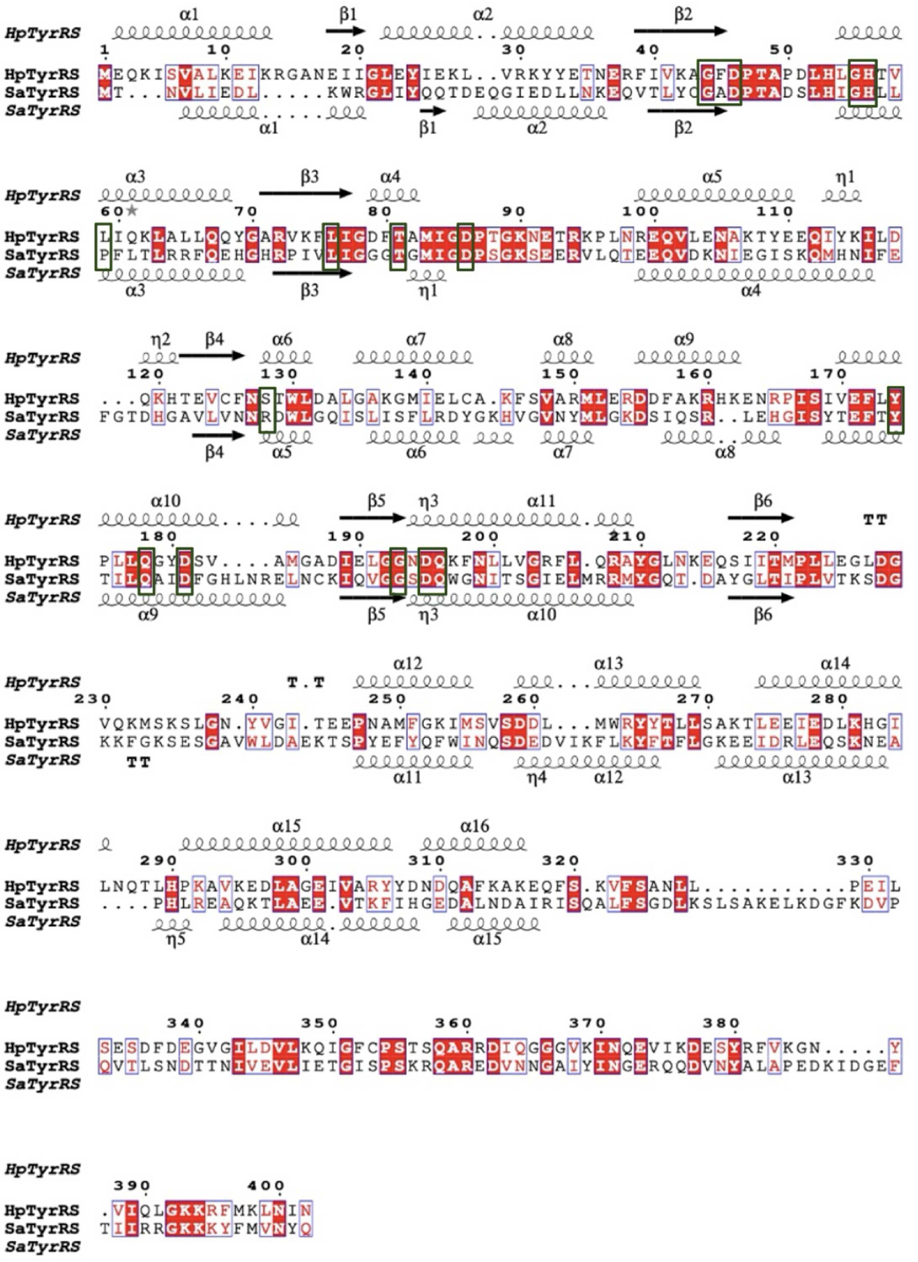
Figure 3
Structure-based primary-sequence alignment of HpTyrRS (PDB entry 6byq) and SaTyrRS (PDB entry 1jii). The secondary-structure elements are as follows: α-helices are shown as large coils, 310-helices as small coils labeled η and β-strands as arrows labeled β, with β-turns labeled TT. Identical residues are shown in white font on a red background, while conserved residues are shown in red font and conserved regions are shown in blue boxes. The residues interacting with SB-219383 are indicated with green boxes. This figure was generated using ESPript 3.0 with alignment in Clustal Omega. |
| STRUCTURAL BIOLOGY COMMUNICATIONS |
ISSN: 2053-230X
Open  access