

 menu
|
|
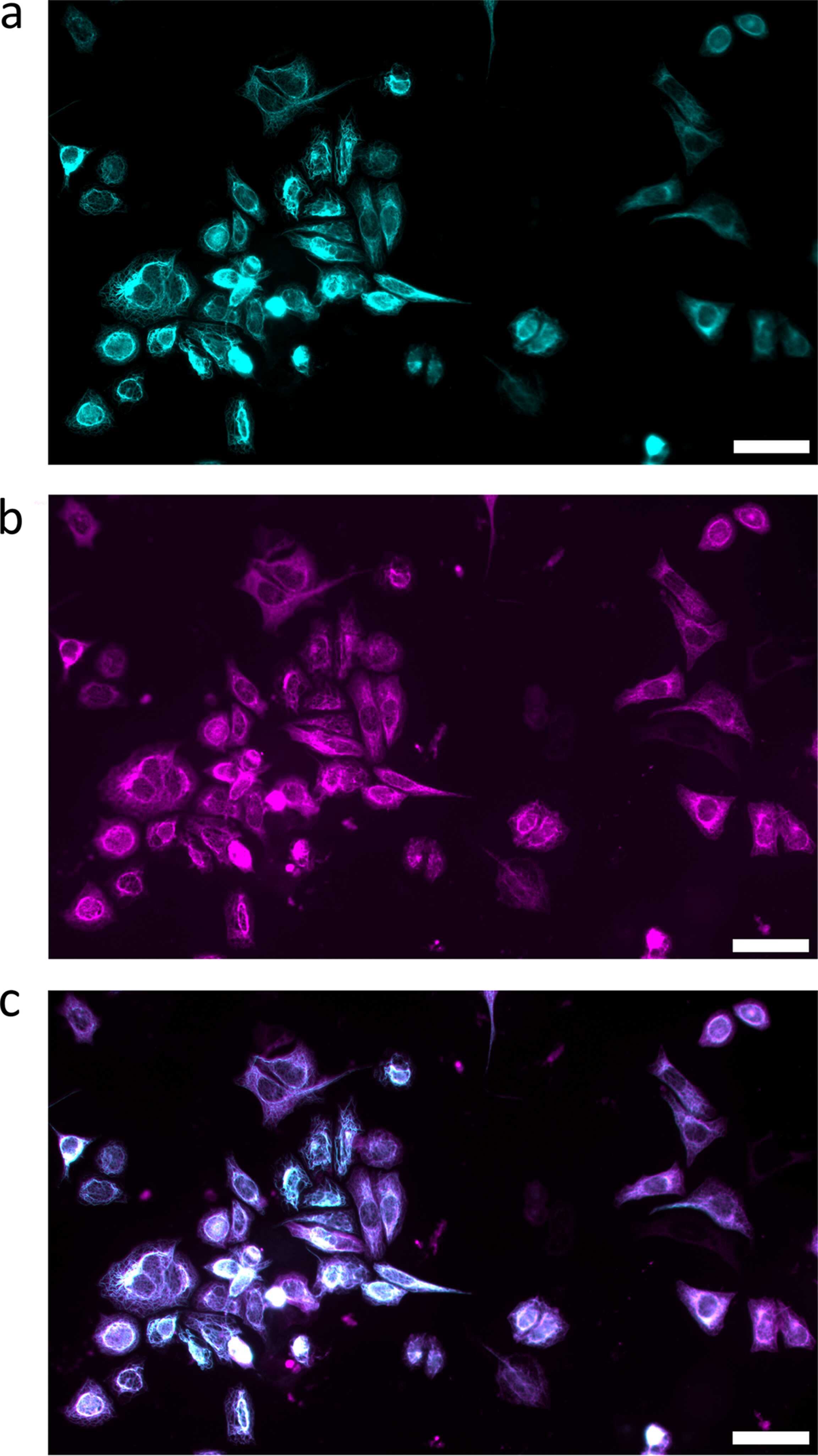
Figure 3
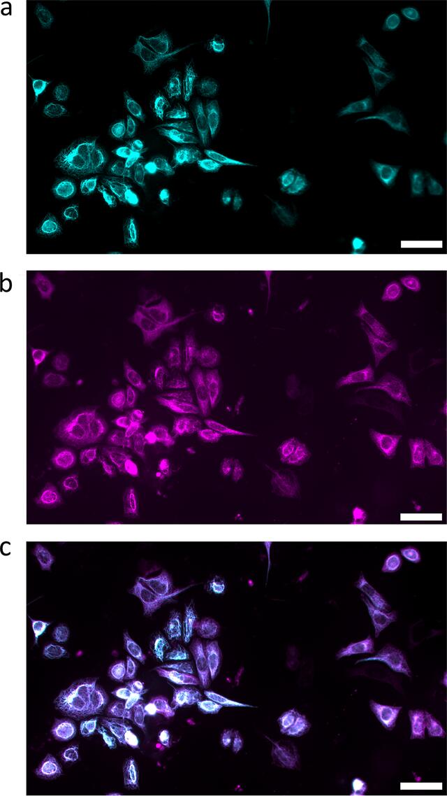
Fluorescence images of fixed-hydrated keratin-rich SW-13 cells before (a, cyan) and after (b, magenta) in situ immunostaining inside the assembled device. The overlay (c) of the keratin structures before and after the in situ immunostaining shows good agreement. Scale bars: 50 µm. |
 | JOURNAL OF SYNCHROTRON RADIATION |
ISSN: 1600-5775
 access