

 menu
|
|
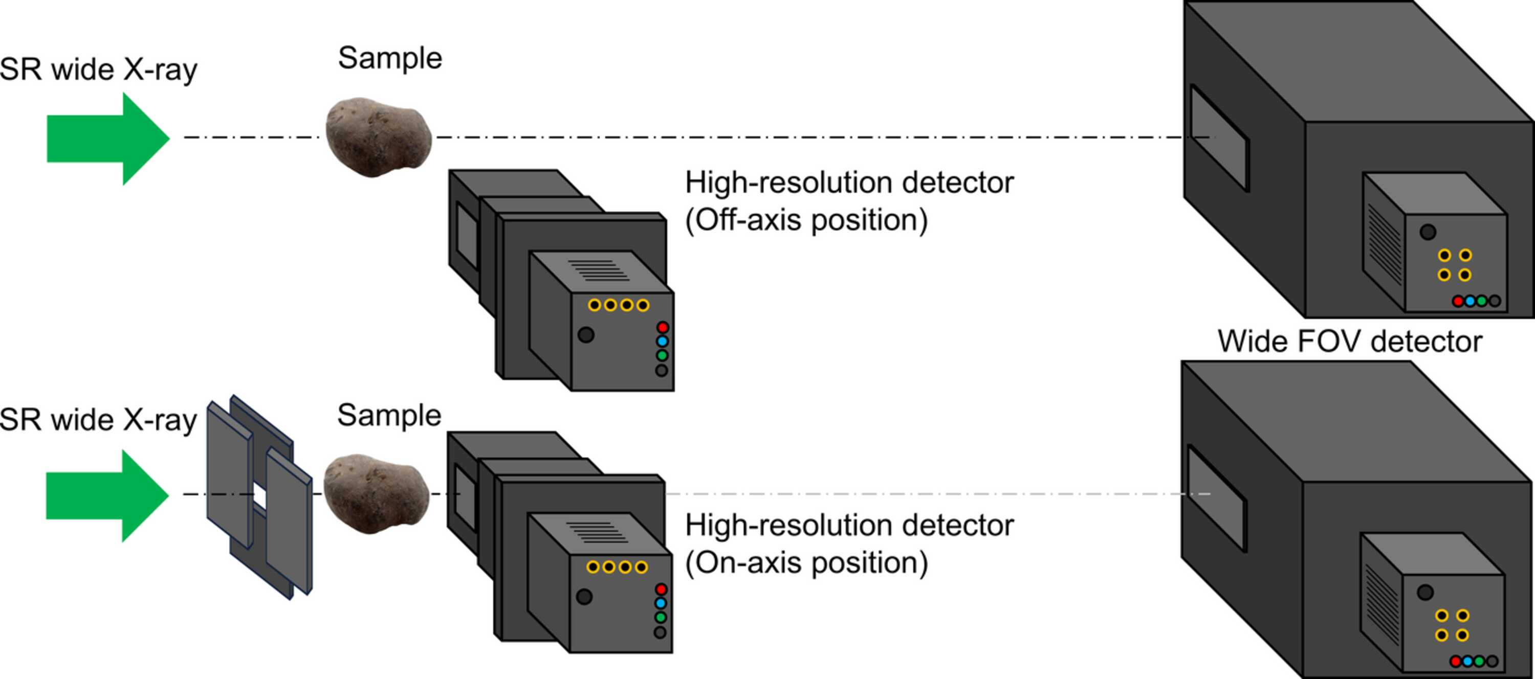
Figure 3
Schematic drawing of the basic concept of X-ray multi-scale imaging comprising a wide-FOV detector and a high-resolution detector; wide-FOV observation (top) and local high-resolution observation (bottom). In the high-resolution observation, a four-quadrant slit was set in front of the sample to avoid irradiation of the wide X-ray beam onto the high-resolution detector. |
 | JOURNAL OF SYNCHROTRON RADIATION |
ISSN: 1600-5775
 access