

 menu
|
|
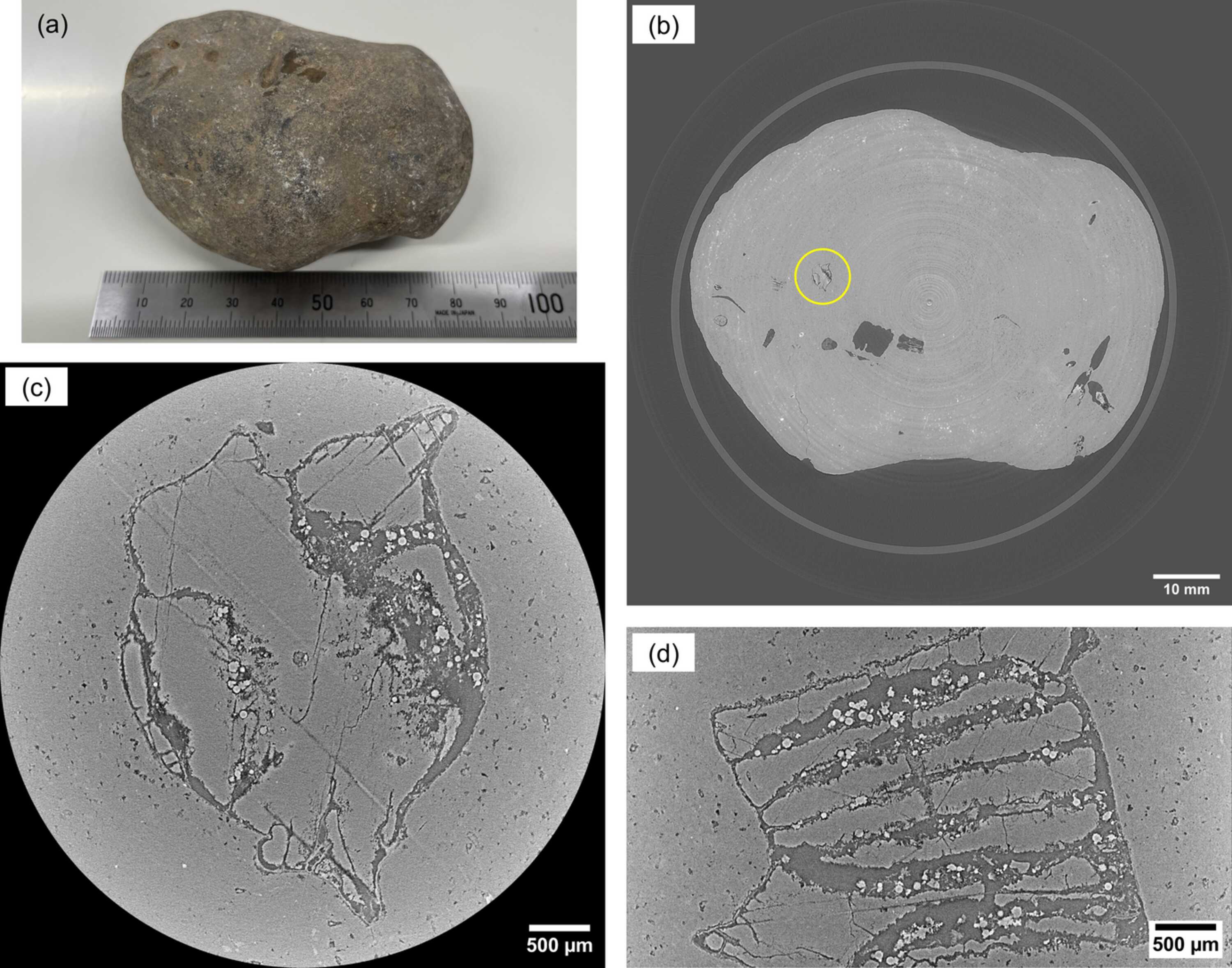
Figure 4
X-ray multi-scale micro-tomography of a nodule. (a) Photograph of the nodule. (b) Wide-FOV sectional image of the entire structure of the nodule. (c) Local high-resolution sectional image at the circle shown in (b). (d) Sectional image perpendicular to the image shown in (c). |
 | JOURNAL OF SYNCHROTRON RADIATION |
ISSN: 1600-5775
 access