

 menu
|
|
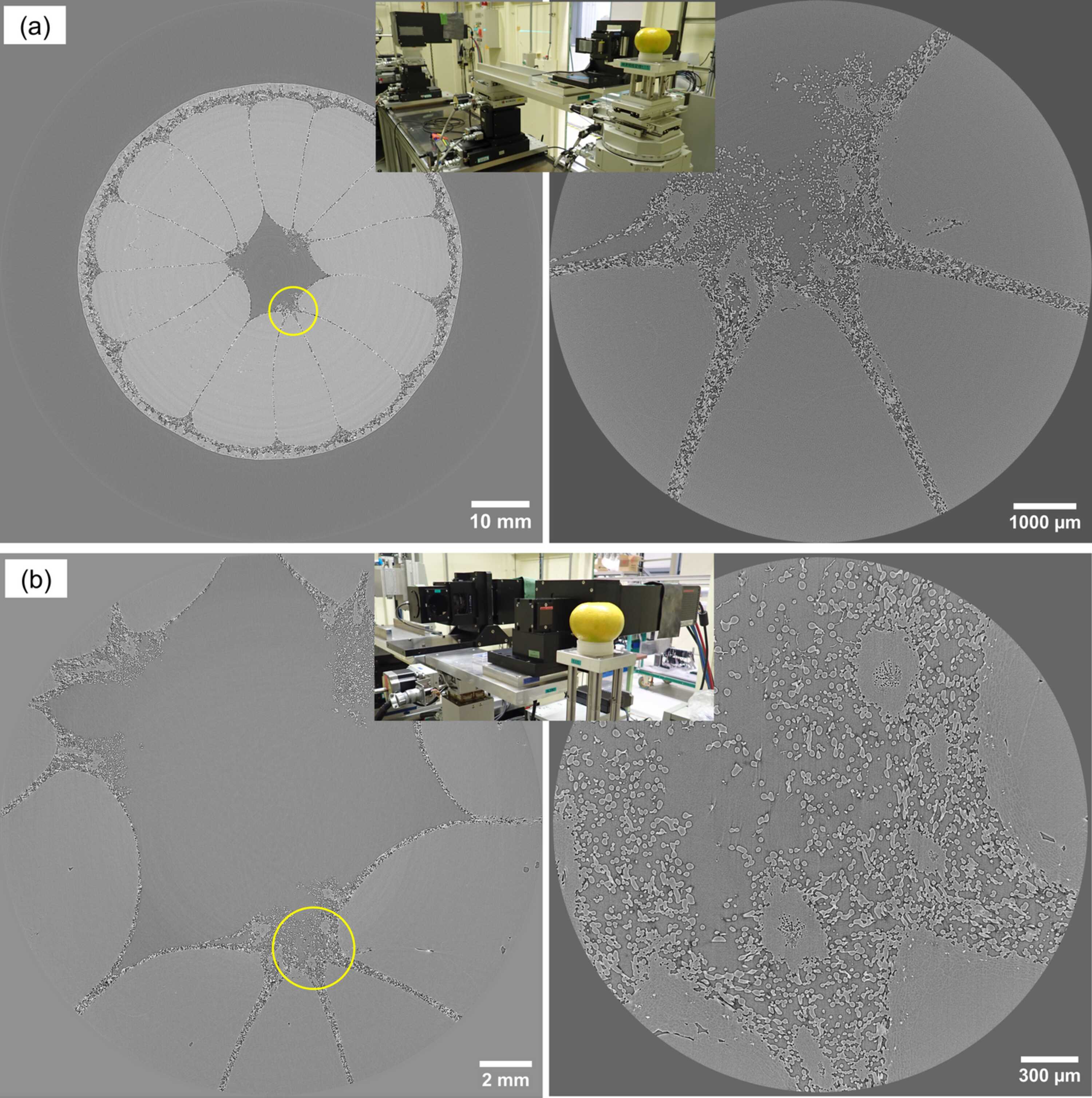
Figure 6
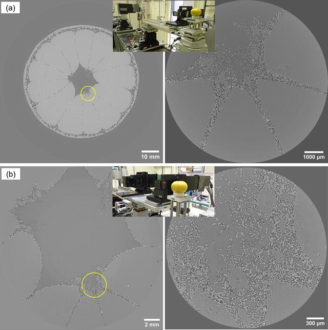
Wide-range X-ray multi-scale micro-tomography of a mandarin orange. (a) Wide-FOV sectional image (left) and local high-resolution sectional image at the circle (right) observed at EH2&3. (b) Wide-FOV sectional image (left) and local high-resolution sectional image at the circle (right) observed at EH1. Insets show photographs of the actual setups for the multi-scale observations. |
 | JOURNAL OF SYNCHROTRON RADIATION |
ISSN: 1600-5775
 access