

 menu
|
|
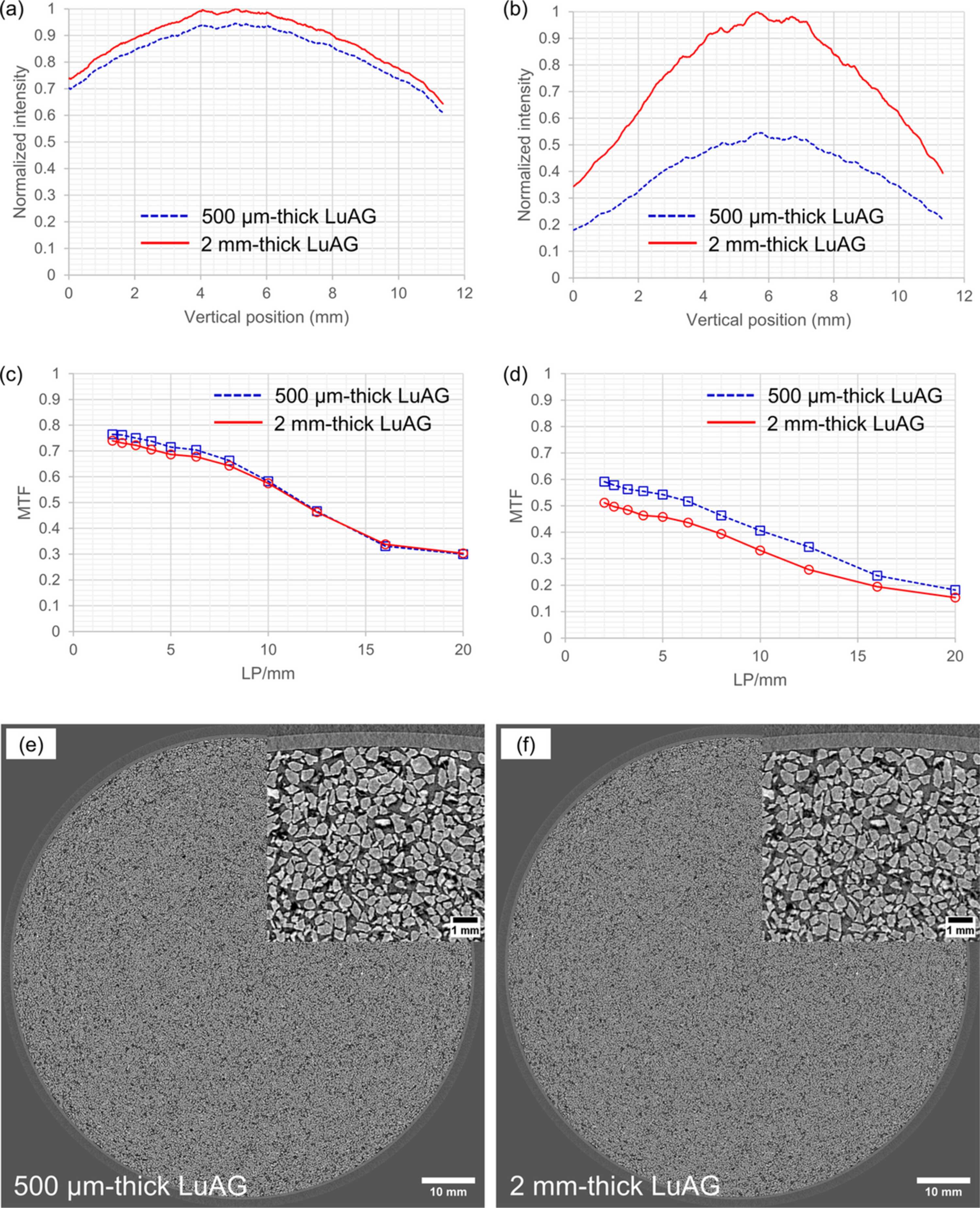
Figure 9
Vertical intensity profiles of (a) 40 keV and (b) 110 keV X-ray beams, measured with the developed wide-FOV X-ray imaging detector. Modulation transfer functions of the wide-FOV X-ray imaging detector, measured with (c) 40 keV and (d) 110 keV X-ray beams. Reconstructed sectional images of silica sand filling a petri dish, observed with (e) 500 µm-thick and (f) 2 mm-thick LuAG crystals. |
 | JOURNAL OF SYNCHROTRON RADIATION |
ISSN: 1600-5775
 access