

 menu
|
|
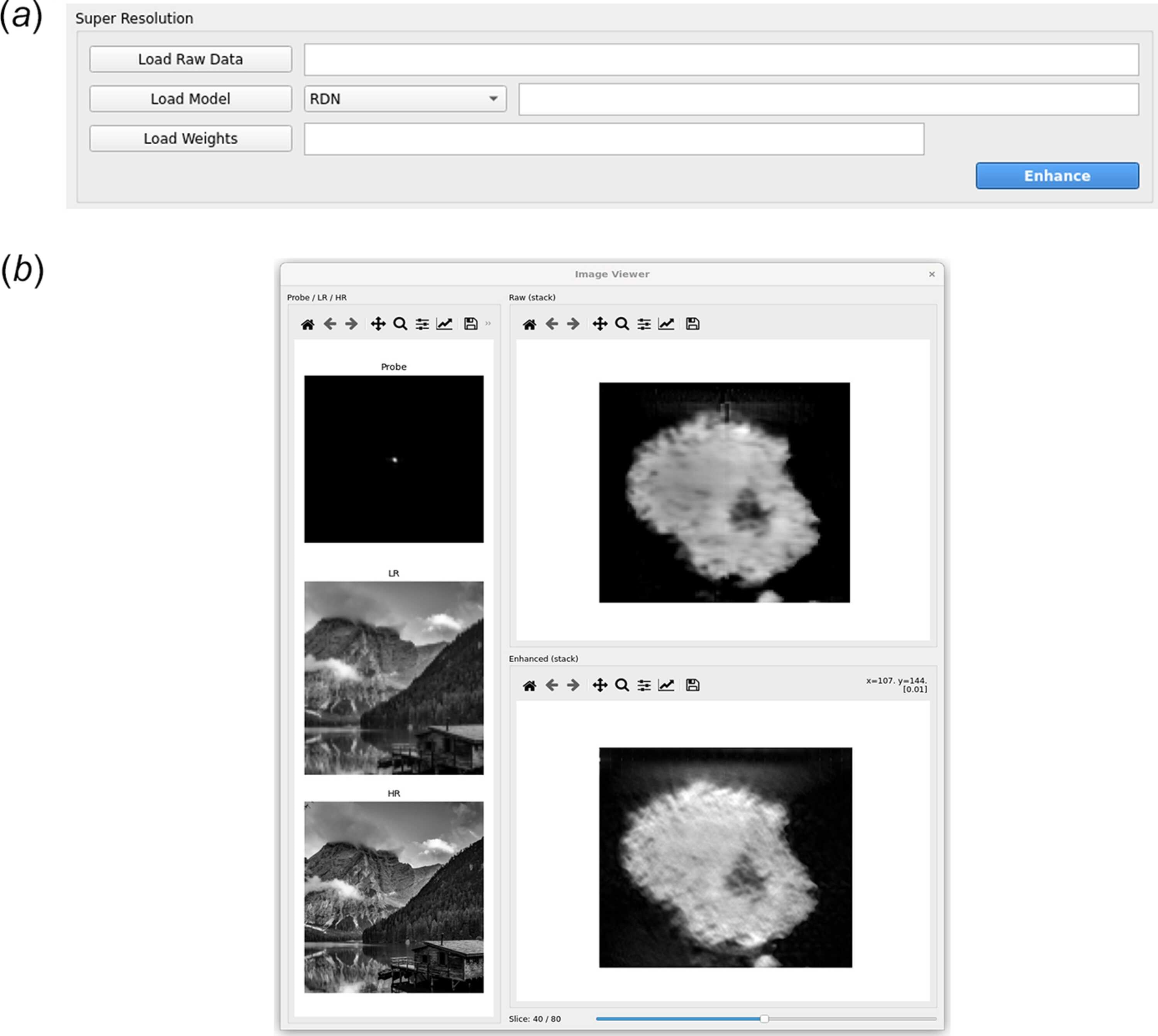
Figure 4
Overview of the GUI for X-ray image enhancement, illustrated using DPC data. (a) The super-resolution panel provides controls for raw data import, model selection, trained weight loading, and execution of the enhancement. (b) The shared visualization window displays the probe and the natural-image LR–HR reference pair retained from the optional preprocessing simulation on the left, and the raw and enhanced DPC images of an NMC cathode particle on the right. The reference pair illustrates the simulated degradation and is not generated during inference on the experimental data. The slice slider appears only for image-stack inputs and allows browsing through individual two-dimensional frames. |
 | JOURNAL OF SYNCHROTRON RADIATION |
ISSN: 1600-5775
 access