journal menu


issue contents
March 1995 issue
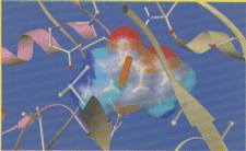
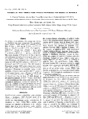
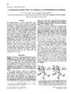
Cover illustration: Active site of purine nucleoside phosphorylase with the electrostatic potential surface and dipole of guanine. Rendered with Ribbons++ and SPARTAN.
research papers


This paper exploits the power of certain 4 x 4 matrices to show how cluster pairs may be selected from an ensemble of clusters of structures and united by superposition, thus developing a tree of related structures, such that each union so formed is the most compact available, All residuals and orientations are available at every stage, and coordinate transformations and average structures are not involved.
A geometric analysis suggests that in certain nucleic acid structures C-H⋯O hydrogen-bonding interactions may occur. These interactions could stabilize the base-base associations.


It was shown that the hexameric structure of an enzyme-inhibitor complex can be determined by molecular replacement using the structure of monomeric native protein as a probe molecule.
PDB reference: 1ayp
The crystal structure of the ligand-binding domain of the Escherichia coli aspartate receptor was determined and compared to the analogous structures from Salmonella typhimurium.
PDB reference: 2asr
The crystal structure of a two iron-sulfur cluster protein the ferredoxin from Clostridium acidurici, has been solved from diffraction data up to 1.8 Å resolution. The refinement was performed by a molecular dynamic technique using the variable force constants.
A simple procedure for obtaining interpretable electron-density maps for crystals where every reflection is exactly overlapped by one other, is presented. The method uses the redundancy inherent in a system possessing noncrystallographic symmetry to provide constraints enabling deconvolution of the data.
The crystal structure of azurin mutant Phe114Ala from P. aeruginosa has been solved to a final crystallographic R value of 18.5% to 2.6 Å resolution. The mutation was performed at residue Phe114 to investigate its suggested role in the electron self-exchange reaction.
PDB reference: 1azn
The structure of narbonin, a seed globulin with (α/β)8-topology, has been refined using the cDNA sequence and compared with the X-ray sequence which was reported earlier. The classification of narbonin as a storage protein without enzymatic function is discussed.
PDB reference: 1nar
Netropsin binding to the B-DNA dodecamer d(CGCGTTAACGCG) causes very little structural perturbation in the DNA.
The structure of a new alkaline serine protease, M-protease, was determined by molecular replacement using the coordinates of subtilisin Carlsberg, and refined to a crystallographic R factor of 0.189 at 2.4 Å resolution.
PDB reference: 1mpt
Large single crystals of hen egg-white lysozyme and the determination of the temperature-dependence of their solubility allowed a new method to be used for the basic physicochemical understanding of the growth processes of protein crystals.
The crystal structure of a Phe→Leu mutation in the hydrophobic core of barnase reveals that the overall structural changes are minimal and that the side-chain conformation of the mutated residue is determined by conformational energy as well as the amount of hydrophobic surface area that is buried by that residue.
PDB reference: 1brg
Preliminary crystal analysis of cytochrome c6, an electron carrier between chlorophyl molecule P700 of PSI and cytochrome f from the b6f complex.
short communications
Bovine ubiquinol cytochrome c oxidoreductase has been crystallized in a new form. The space group is C2221 with unit-cell parameters a = 384, b = 118 and c = 177 Å.
The NAD(P)-dependent GDH from the hyperthermophile P. furiosus has been crystallized by the hanging-drop method of vapour diffusion using lithium sulfate as a precipitant. The determination of the structure of this enzyme will be important in advancing our understanding of how proteins are adapted to enable them to survive at such extreme temperatures.
Single crystals of NAD-dependent GAPDH from E. coli have been obtained. They belonged to space group C2221 with cell dimensions a = 79.1, b = 189.6 and c = 122.2 Å.
PDB reference: 1asi
Single crystals of NAD-dependent GAPDH from E. coli have been obtained. They belonged to space group C2221 with cell dimensions a = 79.1, b = 189.6 and c = 122.2 Å.
book reviews
Free