issue contents

Journal logoBIOLOGICAL
CRYSTALLOGRAPHY
ISSN: 1399-0047

March 2005 issue

Highlighted illustration

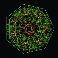
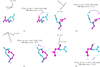
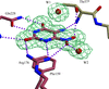
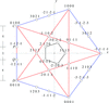
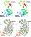
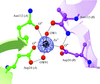
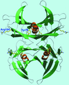
Cover illustration: The double heptameric rings of the PA-Sm1 core protein in complex with RNA are shown in an axial view with the two RNA binding domains, between the heptamers and in their central channel, respectively (p. 269). The hetagonal prismatic forms encasing the three subsystems have heights and radii related by heptagrammal scalings and are expressible in terms of a single parameter u. In particular, the Sm1 core subsystem has a radius re = 8u, an inter-ring distance d= 4u (double the value in the free state) and a total height H = 18u. The corresponding values for the RNA subsystem between the two heptamers are re' = 8u' and H' = 5u' where u' is scaled by the heptagrammal factor [mu] = 0.8629 with respect to u. For the RNA located within the central cavity one has H' ' = 9u'. The Ca2+ ions (filled cyan circles) are located at the vertices of a heptagon in heptagrammal scaling in relation to the envelope as a whole.

research papers


link to html
It has been shown that a high magnetic field of 10 T improves the quality of lysozyme crystals as quantified by comparison of the X-ray diffraction of crystals grown in the presence and absence of a magnetic field of 10 T, from structure comparisons and from molecular-modelling studies with conformational energy searching, from analysis of the anisotropic B factors and finally from analysis of the mosaicity. It is concluded that magnetic enhancement in protein crystal perfection is an important material engineering tool in the study of protein structure and function.


link to html
The structure of the complex of E. coli glutamate decarboxylase (GADα) with the substrate analogue glutarate has been solved at 2.05 Å resolution.

link to html
Runt-domain (RD) proteins regulate transcription of target genes. The DNA duplex TCTGCGGTC/TGACCGCAG, incorporating the binding site for the RD transcription factors (bold), was crystallized in space group P43. X-ray analysis of two crystals diffracting to 1.7 and 2.0 Å resolution, which had slight variations in their unit-cell parameters, revealed two distinct conformations of the A-­DNA helix.

link to html
The architectural elements of axial-symmetric proteins, such as envelopes and channels, are characterized by lattices with a single lattice parameter and by crystallographic scale rotations.

link to html
The architectural elements of axial-symmetric proteins, such as envelopes and channels, are characterized by polygonal lattices with a single lattice parameter and by crystallographic scale rotations.

link to html
The interplay between structural elements in axial-symmetric protein–DNA (or protein–RNA) complexes is shown to be compatible with a crystallographic characterization in terms of integral polygonal lattices and scale-rotational point-group transformations.


link to html
A modified molecular-replacement method is described that makes use of six-dimensional searches and the phased translation function, providing a systematic examination of all possible search-model orientations in an experimental electron-density map.

link to html
The structure of the type I L-asparaginase from the hyperthermophilic archaeon Pyrococcus horikoshii was solved by the MAD method and was refined to 2.16 Å resolution.

link to html
The crystal structure of the zinc-induced heterodimer of two closely related isoforms of phospholipase A2 (PLA2) from cobra venom has been determined and refined at 2.7 Å resolution. The structure reveals that zinc ion is unable to bind in the so-called calcium-binding loop of PLA2 and that by inducing dimerization zinc ion inactivates the two isoforms.

link to html
The crystal structure of the cofactor-dependent phosphoglycerate mutase of M. tuberculosis has been determined at a resolution of 1.70 Å.

link to html
The X-ray structure of recombinant human cyclophilin J, a novel member of the cyclophilin family, has been determined at 2.6 Å resolution. The structure is similar to the cyclophilin A structure and appears to support the solvent-assisted mechanism.

link to html
Energy restraints derived from linear-scaling semiempirical quantum-mechanical calculations and X-ray diffraction data are combined to refine the crystal structure of a protein molecule, bovine pancreatic trypsin inhibitor.

link to html
The crystal structures of two transthyretin variants TTR Y78F, an amyloidogenic protein, and TTR R104H, a non-pathogenic/protective variant, have been determined. The structural features responsible for TTR stabilization/destabilization are discussed.

short communications


link to html
A cryoprotection scheme was developed that results in glycerol binding within the interior of the membrane-spanning domain. The His-tagged recombinant protein adopts different lattice contacts but diffracts as well as the native protein.

link to html
The structure determination of the major outer surface antigen of the B. anthracis spore using automated phasing programs and exploiting the anomalous signal from iodide is presented.

link to html
Pseudo-merohedrally twinned crystals of an ADP-forming acetyl coenzyme A synthetase from P. furiosus have been produced.
Follow Acta Cryst. D
Sign up for e-alerts
Follow Acta Cryst. on Twitter
Follow us on facebook
Sign up for RSS feeds