journal menu


issue contents
October 2014 issue
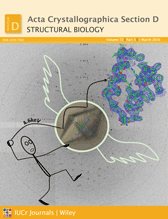
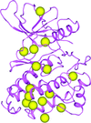
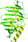
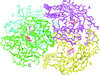
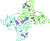
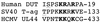
Cover illustration: Allophycocyanin B from Synechocystis PCC 6803 (p. 2558). Eight ![[alpha]](/logos/entities/alpha_rmgif.gif) /
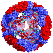
/![[beta]](/logos/entities/beta_rmgif.gif) trimers form a spherical assembly in the crystal lattice. subunits (ApcD) are coloured blue, subunits (ApcB) red and the PCB chromophore is cyan. One of the eight trimers is represented as a ribbon diagram.
trimers form a spherical assembly in the crystal lattice. subunits (ApcD) are coloured blue, subunits (ApcB) red and the PCB chromophore is cyan. One of the eight trimers is represented as a ribbon diagram.
diffraction data deposition


Free 
An introduction to a collection of articles from the IUCr Diffraction Data Deposition Working Group.

Open access
access
A local raw `diffraction data images' archive was made available and some data sets were retrieved and reprocessed, which led to analysis of the anomalous difference densities of two partially occupied Cl atoms in cisplatin as well as a re-evaluation of the resolution cutoff in these diffraction data. General questions on storing raw data are discussed. It is also demonstrated that often one needs unambiguous prior knowledge to read the (binary) detector format and the setup of goniometer geometries.
Open access
access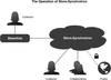
The Store.Synchrotron service, a fully functional, cloud computing-based solution to raw X-ray data archiving and dissemination at the Australian Synchrotron, is described.
Open access
access
An analysis is performed of the technical and financial challenges to be overcome if deposition of primary experimental data is to become routine.
Open access
access
Macromolecular structures deposited in the PDB can and should be continually reinterpreted and improved on the basis of their accompanying experimental X-ray data, exploiting the steady progress in methods and software that the deposition of such data into the PDB on a massive scale has made possible.
research papers

Anomalous diffraction signals may be routinely enhanced for native-SAD analysis by merging multi-crystal data sets at 6 keV.
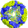
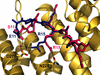
In the crystal lattice, eight trimers of allophycocyanin B form a porous spherical, 48-subunit assembly, in which the phycocyanobilin chromophore is co-planar.
PDB reference: allophycocyanin B, 4po5
Open access
access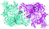
Details of the RNA polymerase I crystal structure determination provide a framework for solution of the structures of other multi-subunit complexes. Simple crystallographic experiments are described to extract relevant biological information such as the location of the enzyme active site.
Cthe_2159 is a member of a domain of unknown function family (DUF4353) and was identified from the C. thermocellum genome by its ability to bind cellulose. The structure was determined using gadolinium SAD phasing and shows structural features that are highly similar to some polysaccharide lyase families.
Open access
access
Rank scaling of Fourier syntheses leads to new tools for the comparison of crystallographic contour maps. The new metrics are in better agreement with a visual map analysis than the conventional map correlation coefficient.
The thermostability of the α-carbonic anhydrase from the thermophilic bacterium T. ammonificans is shown to depend on the formation of intrasubunit disulfide bonds and a unique intersubunit disulfide/lysine core at the centre of the tetrameric molecule. Unlike most α-carbonic anhydrases, this enzyme does not show esterase activity towards p-nitrophenyl acetate, which is proposed to be owing to its increased structural rigidity.
The crystal structure of the lipoprotein LprF found in M. tuberculosis and M. bovis has been determined at 1.1 Å resolution.
PDB reference: LprF, 4qa8
Open access
access
Peptidoglycan O-acetylesterase (Ape1), which is required for host survival in Neisseria sp., belongs to the diverse SGNH hydrolase superfamily, which includes important viral and bacterial virulence factors. Here, multi-domain crystal structures of Ape1 with an SGNH catalytic domain and a newly identified putative peptidoglycan-detection module are reported.
The crystal structure of arabinose-5-phosphate isomerase from B. fragilis NCTC 9343 in complex with an endogenous ligand, CMP-Kdo, is described. Structural and sequence comparisons suggest highly conserved functional residues in the active site that could be targeted for antibacterial drug design.
PDB reference: arabinose-5-phosphate isomerase, 3etn
Open access
access
A new indexing method is presented which is capable of indexing multiple crystal lattices from narrow wedges of data. The efficacy of this method is demonstrated with both semi-synthetic multi-lattice data and real multi-lattice data recorded from microcrystals of ∼1 µm in size.
A new crystal structure of the human nNOS heme domain and a better diffracting crystal form of human eNOS heme domain are described.
It is shown that even a low dose (0.06 MGy) of synchrotron radiation absorbed by a protein crystal induces structural alterations. This finding raises a significant issue as it may question previously solved structures as well as highlighting and defining new limitations of protein X-ray crystallography.

A case study on the treatment of SIRAS data using the originally unknown protein LegC3N is described. An iterative direct-method-based treatment was proposed that led to improved results in this particular test case.
Open access
access
The same hexagonal array of archaerhodpsin-2 trimers as observed in the native membrane is reconstituted in a three-dimensional crystal prepared by the membrane fusion method. In this crystal, a pair of conserved glutamate residues in the proton-release pathway is maintained by a low-barrier hydrogen bond.
PDB reference: archaerhodopsin-2, 3wqj
Open access
access
A robust and transferable algorithm is presented to objectively describe and rank robotically captured images of crystallization droplets according to their likelihood of crystalline behaviour for the efficient and accurate identification of successful crystallization.
High-multiplicity data were used to solve the West Nile virus NS1 structure by S-SAD and for the extension of useable resolution for refinement.
PDB reference: West Nile virus NS1, 4tpl
The C. jejuni phosphoethanolamine transferase EptC functions in antibacterial resistance, motility and host intestinal colonization. In this study, the three-dimensional structure of the catalytic domain of EptC is reported in a covalent phosphoenzyme-intermediate state. EptC active-site mutant strains of C. jejuni demonstrate the potential for EptC enzyme inhibitors.
PDB reference: EptC, 4tn0
Open access
access
The high-resolution crystal structures of the human tankyrase 2 poly(ADP-ribose) polymerase (PARP) domain in complex with 16 various PARP inhibitors are reported, including the compounds BSI-201, AZD-2281 and ABT-888, which are currently in Phase 2 or 3 clinical trials.
PDB references: TNKS2–EB-47, 4tk5; TNKS2–DR-2313, 4pnl; TNKS2–3,4-CPQ-5-C, 4tju; TNKS2–BSI-201, 4tki; TNKS2–TIQ-A, 4pnr; TNKS2–5-AIQ, 4pnq; TNKS2–4-HQN, 4pnn; TNKS2–3-AB, 4pml; TNKS2–AZD-2281, 4tkg; TNKS2–NU-1025, 4pnm; TNKS2–PJ-34, 4tjw; TNKS2–INH2BP, 4pns; TNKS2–DPQ, 4tk0; TNKS2–ABT-888, 4tjy; TNKS2–IWR-1, 4tkf; TNKS2–1,5-IQD, 4pnt
The X-ray structure of the N-terminal acetyltransferase domain [AAC(6′)-Ie] of the bifunctional antibiotic-resistance enzyme AAC(6′)-Ie-APH(2′′)-Ia has been determined as a ternary kanamycin/coenzyme A complex. X-ray structures of the isolated AAC(6′)-Ie and APH(2′′)-Ia domains, combined with small-angle X-ray scattering (SAXS) data, allowed the determination of the structural architecture of the full-length bifunctional enzyme in solution.
PDB reference: AAC(6′)-Ie, 4qc6
Open access
access
[AsnB26]- and [GlyB26]-insulin mutants attain a B26-turn like fold without assistance of chemical modifications. Their structures match the insulin receptor interface and expand the spectrum of insulin conformations.
letters to the editor
Free
Free
A response to the article by Alvisi & Jans [(2014), Acta Cryst. D70, 2775_2776].
Free
Updating literature references in the Protein Data Bank is discussed.
Free
A response to the article On the prompt update of literature references in the Protein Data Bank [Wlodawer (2014), Acta Cryst. D70, 2779].