journal menu


issue contents
 | JOURNAL OF APPLIED CRYSTALLOGRAPHY |
ISSN: 1600-5767
X-ray diffraction microscopy special issue (March 2013)
Guest Editor(s): Andras Borbely
This virtual issue on recent developments in X-ray diffraction microscopy collects together a series of articles originally published in the journal between August 2012 and April 2013. These articles focus on novel diffraction methods that enable visualization of the internal structure of crystalline materials from the millimetre down to the nanometre scale.
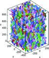
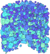
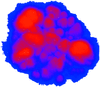
Cover illustration: Structures of polycrystals and of a Schizosaccharomyces pombe spore at different length scales. Courtesy of Reischig et al. [J. Appl. Cryst. (2013), 46, 297–311], Larson & Levine [J. Appl. Cryst. (2013), 46, 153–164] and Rodriguez et al. [J. Appl. Cryst. (2013), 46, 312–318].


Free 
In anticipation of the International Year of Crystallography in 2014, Journal of Applied Crystallography presents a virtual issue on recent developments in X-ray diffraction microscopy. This issue is available at //journals.iucr.org/special_issues/2013/imaging/.
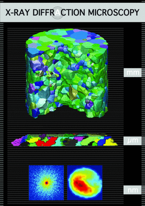
Three-dimensional X-ray diffraction microscopy is a fast and nondestructive structural characterization technique aimed at studies of the individual crystalline elements (grains or subgrains) within millimetre-sized polycrystalline specimens. A review of the field is provided, with a viewpoint from materials science.
Recent developments in the use of the diffraction contrast tomography method to create three-dimensional grain maps of polycrystalline samples are described. These include the use of detectors in arbitrary positions, including high Bragg angles, and the application to multiphase materials.
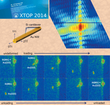
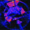
The evolution of the crystallographic orientation field in a polycrystalline sample of copper is mapped in three dimensions as tensile strain is applied. Using forward-modeling analysis of high-energy X-ray diffraction microscopy data, the ability to track intragranular orientation variations is demonstrated.
The underlying techniques and capabilities of submicrometre-resolution three-dimensional X-ray microscopy (3DXM) are reviewed. The major applications and accomplishments of 3DXM are discussed through selected examples, including investigations of local elastic strain, plastic strain, crystal structure, phase changes and microstructure evolution.
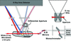
Combining diffraction/scattering and tomography methods allows the three-dimensional structural imaging of heterogeneous samples to discriminate multiple crystalline and amorphous materials. The principle and applications of the methods are described and the reconstruction artefacts and limitations are discussed.
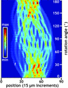
The microstructure of an SiC-monofilament-reinforced Al matrix is reconstructed with 15 µm in-plane volume elements using microcomputed tomography based on diffracted intensities from SiC and Al phases.
Oversampling smoothness: an effective algorithm for phase retrieval of noisy diffraction intensities
An iterative phase retrieval algorithm, termed oversampling smoothness (OSS), has been developed to reconstruct fine features in weakly scattered objects such as biological specimens from noisy experimental data. OSS is expected to find application in the rapidly growing coherent diffraction imaging field as well as other disciplines where phase retrieval from noisy Fourier magnitudes is needed.
Open access
access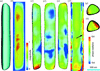
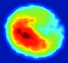
A complex three-dimensional quantitative image of an extended zinc oxide crystal has been obtained using Bragg coherent diffraction imaging integrated with ptychography.