
Acta Crystallographica
Section E CRYSTALLOGRAPHIC COMMUNICATIONS


 menu


research communications
| CRYSTALLOGRAPHIC COMMUNICATIONS |
 access
and Hirshfeld surface analysis of 6-methyl-2,3-dihydro-1,3-benzothiazol-2-iminium 3-(1,3-benzothiazol-2-ylsulfanyl)propanoate

aTermez State University, Barkamol Avlod Str. 43, Termez 190111, Uzbekistan, and bInstitute of Bioorganic Chemistry, Academy of Sciences of Uzbekistan, 100125, M. Ulugbek Str 83, Tashkent, Uzbekistan
*Correspondence e-mail: [email protected]
In the title salt, C8H9N2S+·C10H8NO2S2−, formed by proton transfer from the carboxylic acid to the ring N atom of 6-methyl-1,3-benzothiazol-2-amine, a 6-methyl-2,3-dihydro-1,3-benzothiazol-2-iminium cation (A) and a 3-(1,3-benzothiazol-2-ylsulfanyl)propanoate anion (B) are present. Deprotonation of the carboxyl group is evidenced by its two distinctly different C—O bond lengths of 1.281 (6) and 1.220 (6) Å, while protonation of the ring N atom is confirmed by the localization of the corresponding H atom in a difference-Fourier map. Despite their different ionic states, the benzothiazole fragments of the two components retain closely comparable bond lengths and angles. The cation and anion are linked by three charge-assisted N—H⋯O hydrogen bonds. Two of these, involving the ring N—H group and one of the amino N—H groups, bridge the two carboxylate O atoms of a single anion to form an R22(8) graph-set motif; the third links these ion pairs into layers parallel to (100). Hirshfeld surface analysis, performed separately for components A and B, shows that H⋯H contacts dominate the surface for A (38.9%), followed by C⋯H/H⋯C (24.2%), O⋯H/H⋯O (13.0%) and S⋯H/H⋯S (11.9%) contacts. For anion B, the principal contributions are H⋯H (27.5%), S⋯H/H⋯S (25.7%), C⋯H/H⋯C (17.2%) and O⋯H/H⋯O (15.3%). The complementary H⋯O/O⋯H contributions are consistent with the important role of the charge-assisted N—H⋯O hydrogen bonds in consolidating the crystal packing.
Keywords: crystal structure; organic salt; benzothiazol-3-ium; proton transfer; charge-assisted hydrogen bonding.
CCDC reference: 2515049
1. Chemical context
Benzothiazole is an aromatic heterobicyclic scaffold formed by fusion of a benzene ring with a thiazole ring, combining an endocyclic N atom and an S atom within a single rigid framework (Padilla-Martínez et al., 2025 ). This dual-heteroatom arrangement gives the core exceptional versatility as a hydrogen-bonding acceptor and as a metal-coordinating site, while substitution at the 2-position allows fine control over its donor⋯acceptor character, making benzothiazole an extensively exploited heterocycle in medicinal and supramolecular chemistry (Yadav et al., 2023). Benzothiazole derivatives display a remarkably broad spectrum of biological activities, including antimicrobial, antitumour, anti-inflammatory, antidiabetic, antiviral and anti-Alzheimer's properties (Yadav et al., 2023; Al-Mutairi, 2023), and several of these compounds are established clinical agents, among them riluzole (amyotrophic lateral sclerosis), ethoxzolamide (glaucoma), frentizole (immunosuppressive) and the amyloid-imaging probe thioflavin-T (Padilla-Martínez et al., 2025). Beyond medicinal chemistry, benzothiazole derivatives are of considerable practical importance as rubber-vulcanization accelerators, corrosion inhibitors, dyes and agrochemical intermediates.

In 2-aminobenzothiazoles, the endocyclic N atom can act as a proton acceptor, while the exocyclic amino group provides additional hydrogen-bonding donor sites. Conversely, S-functionalized derivatives of 2-mercaptobenzothiazole combine a polarizable thioether linkage with other functional groups such as carboxylic acids.
Salts formed by proton-transfer between carboxylic acids and heterocyclic bases are particularly useful for crystal-engineering studies because proton transfer creates complementary charged donor and acceptor sites. The resulting N—H+ and COO− groups commonly generate short, charge-assisted N—H⋯O hydrogen bonds and robust supramolecular synthons. Previous structural studies of 2-aminobenzothiazole with carboxylic acids have shown that protonation commonly occurs at the endocyclic N atom and that the ionic components are frequently organized by paired N—H⋯O interactions (Lynch et al., 1998, 1999; Zhang et al., 2009; Ilkimen et al., 2013).
Synthesis and crystal structure determination of benzothiazole-derived carboxylic acids and their salts have also been a continuing subject of our work (Ashurov, 2016; Ashurov et al., 2016, 2017, 2018). Herein, we report the crystal structure and Hirshfeld surface analysis of a new proton-transfer salt formed between 3-(1,3-benzothiazol-2-ylsulfanyl)propanoic acid and 6-methyl-1,3-benzothiazol-2-amine. Single-crystal X-ray diffraction demonstrates complete proton transfer from the carboxyl group to the ring N atom of the aminobenzothiazole component, giving the 1:1 organic salt C8H9N2S+·C10H8NO2S2−.
2. Structural commentary
The of the title salt contains one 6-methyl-2,3-dihydro-1,3-benzothiazol-2-iminium cation, (A, with all atoms of this unit designated by suffix A) and one 3-(1,3-benzothiazol-2-ylsulfanyl)propanoate anion (B, with all atoms of this unit designated by suffix B), Fig. 1. The compound crystallizes in the orthorhombic space group Pna21 with Z = 4.
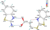 | Figure 1 The asymmetric unit of the title compound, showing the atom-numbering scheme; displacement ellipsoids are drawn at the 50% probability level and H atoms are shown as small spheres of arbitrary radius. N—H⋯O hydrogen bonds forming the R22(8) graph-set motif are shown as blue dashed lines and the intramolecular C9B—H9BA⋯N1B contact as a red dashed line. |
Proton transfer was clearly established by the location of atom H1A at the endocyclic atom N1A in a difference-Fourier map; the exocyclic amino atom N2A carries two H atoms. Within the protonated 2-aminobenzothiazolium fragment, the N1A—C1A and N2A—C1A distances are 1.316 (6) and 1.300 (7) Å, and the N2A—C1A—N1A angle is 124.8 (5)°. The near-equality of the two N—C1A bond lengths indicates substantial delocalization of the positive charge over the N1A—C1A—N2A fragment, a feature typical of protonated amidine-like systems. The cation is essentially planar: all non-H atoms lie within 0.013 Å of its least-squares plane.
The anion contains a benzothiazole ring system attached through the exocyclic sulfanyl atom S2B to a propanoate chain. The N1B—C1B, S1B—C1B and S2B—C1B bond lengths are 1.298 (7), 1.745 (5) and 1.747 (6) Å, respectively, while S2B—C8B is 1.809 (5) Å. The benzothiazole portion of the anion is also essentially planar; inclusion of S2B gives a maximum deviation of 0.037 Å from the least-squares plane, whereas the propanoate chain is markedly displaced from this plane.
The mean planes of the benzothiazolium ring system of the cation and the benzothiazole ring system of the hydrogen-bonded anion within the subtend a dihedral angle of 56.06 (17)°.
The carboxylate group is characterized by C10B—O1B and C10B—O2B distances of 1.281 (6) and 1.220 (6) Å, respectively, and an O2B—C10B—O1B angle of 124.2 (4)°. The unequal C—O distances reflect the different hydrogen-bonding environments of O1B and O2B (Table 1): O1B, which is involved in the longer C—O bond, accepts two N—H⋯O hydrogen bonds, whereas O2B accepts only one.
|
 .
The flexible sulfanylpropanoate substituent adopts a folded conformation relative to the benzothiazole plane. The C1B—S2B—C8B—C9B torsion angle is −73.6 (4)°, whereas the terminal part of the chain is close to anti, with S2B—C8B—C9B—C10B = −174.5 (3)°. The carboxylate group is nearly coplanar with the C8B—C9B—C10B segment [C8B—C9B—C10B—O1B = −175.4 (4)° and C8B—C9B—C10B—O2B = 4.1 (6)°]. The conformation of the chain is accompanied by an intramolecular C9B—H9BA⋯N1B contact (Table 1).
3. Supramolecular features
In the crystal, the N1A—H1A⋯O1B and N2A—H2AB⋯O2B hydrogen bonds (Table 1) link the cation and the anion of the asymmetric unit into an R22(8) graph-set motif (Etter et al., 1990), in which the carboxylate group bridges the two N—H donor groups of the cation (Fig. 1). The third hydrogen bond, N2A—H2AA⋯O1Bi, connects these ion pairs into layers parallel to (100), Fig. 2.
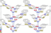 | Figure 2 Part of the crystal structure viewed approximately along the b axis, showing the N—H⋯O hydrogen-bonded layer extending parallel to (100). N—H⋯O hydrogen bonds are shown as blue and C—H⋯N contacts as red dashed lines; H atoms not involved in hydrogen bonding have been omitted for clarity. |
Within these layers, the carboxylate atom O1B is located above the benzothiazolium ring system of a cation related by translation along the b axis, with O1B⋯Cg distances of 3.486 (4), 3.711 (4) and 3.440 (4) Å to the centroids of the thiazolium ring, the phenyl ring and the entire nine-membered ring system, respectively. Neither significant π–π stacking interactions [shortest centroid-to-centroid separation is 4.557 (4) Å] nor C—H⋯π interactions are observed.
4. Hirshfeld surface analysis
A Hirshfeld surface analysis was performed with CrystalExplorer (Spackman et al., 2021) to quantify intermolecular contacts in the crystal packing. The cation (A) and the anion (B) were selected separately, which allows the donor/acceptor environments of the ions to be compared directly with the hydrogen-bonding scheme. The percentage contributions of the individual contacts are summarized in Fig. 3, and the corresponding two-dimensional fingerprint plots are shown in Fig. 4.
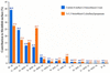 | Figure 3 Percentage contributions of intermolecular contacts to the Hirshfeld surfaces of the 6-methyl-2,3-dihydro-1,3-benzothiazol-2-iminium cation (A) and the 3-(1,3-benzothiazol-2-ylsulfanyl)propanoate anion (B). |
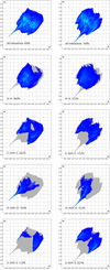 | Figure 4 Two-dimensional fingerprint plots for the cation (left) and anion (right): full, and resolved into H⋯H, C⋯H/H⋯C, O⋯H/H⋯O and S⋯H/H⋯S contacts. |
For cation A, the Hirshfeld surface volume and area are 195.33 Å3 and 194.85 Å2, with a globularity of 0.836 and an asphericity of 0.195 (Fig. 5a). H⋯H contacts make the largest contribution, followed by C⋯H/H⋯C, O⋯H/H⋯O and S⋯H/H⋯S contacts (Figs. 3, 4). Because the cation contains no O atoms, its O⋯H/H⋯O contribution necessarily arises from cationic H atoms in contact with O atoms of neighbouring anions, in agreement with the three N—H⋯O hydrogen bonds, which appear as the most intense red spots on the dnorm surface (Fig. 5a).
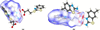 | Figure 5 Hirshfeld surfaces mapped over dnorm for (a) the cation and (b) the anion. Red spots mark close intermolecular contacts corresponding to the N—H⋯O hydrogen bonds. |
For anion B, the volume and area increase to 255.79 Å3 and 248.36 Å2, while the globularity decreases to 0.785 and the asphericity increases slightly to 0.207 (Fig. 5b). Its fingerprint plot is more heterogeneous, with H⋯H and S⋯H/H⋯S contacts making the largest contributions, followed by C⋯H/H⋯C and O⋯H/H⋯O contacts (Figs. 3, 4). The Oinside⋯Houtside contribution of the anion closely matches the complementary Hinside⋯Ooutside contribution of the cation; the two independently calculated surfaces therefore describe opposite sides of the same N—H⋯O contacts.
The larger surface, lower globularity and higher asphericity of the anion are consistent with its additional propanoate chain, and its sulfur-rich surface accounts for the much larger S⋯H/H⋯S contribution compared with the cation (Fig. 3). Overall, the Hirshfeld analysis shows that charge-assisted N—H⋯O hydrogen bonds are the principal directional interactions, supplemented by H⋯H, C⋯H/H⋯C and S⋯H/H⋯S contacts. The percentages represent fractions of the Hirshfeld surface area and therefore indicate contact prevalence rather than interaction strength.
5. Database survey
A search of the Cambridge Structural Database (CSD, Version 6.01, November 2025, including the February 2026 updates; Groom et al., 2016) revealed no structure containing the title ion pair. The discussion below is therefore restricted to the most closely related systems, i.e. salts of 2-aminobenzothiazoles with carboxylic acids and compounds containing the (1,3-benzothiazol-2-ylsulfanyl)alkanoate fragment.
The structure of the neutral base, 6-methyl-1,3-benzothiazol-2-amine, has been reported by Saeed et al. (2007). Protonation of 2-aminobenzothiazoles by carboxylic acids at the endocyclic N atom is well documented. In the series of 2-aminobenzothiazole adducts with carboxylic acids described by Lynch et al. (1998, 1999), a frequently observed association between the base and the acid is the R22(8) graph-set motif formed by the ring N—H and amino N—H donors and the two O atoms of a carboxy(late) group, i.e. the same heterosynthon as found in the title salt. The same cation as in the title compound, 6-methyl-2,3-dihydro-1,3-benzothiazol-2-iminium, has been characterized as a salt with dipicolinic acid (Ilkimen et al., 2013), and further examples of 2-aminobenzothiazolium carboxylates have been described by Zhang et al. (2009), Chen et al. (2020), Gao et al. (2021) and Tsemeugne et al. (2025).
The anion of the title salt is the higher homologue of 2-(1,3-benzothiazol-2-ylsulfanyl)acetate, which has been structurally characterized as the free acid (Fang & Wang, 2010) and as a ligand in CuII and CdII complexes (Bian et al., 2007a,b). Closely related benzothiazole-derived carboxylate salts with amines have also been reported by our group (Ashurov et al., 2016, 2017, 2018; Ashurov, 2016). Compared with the acetate analogues, the additional methylene group of the propanoate chain increases its conformational freedom, which in the title salt allows the carboxylate group to fold away from the benzothiazole plane and to face the donor groups of the cation.
6. Synthesis and crystallization
All starting materials and solvents were obtained from commercial sources and used without further purification. 6-Methyl-1,3-benzothiazol-2-amine (40 mg, 0.244 mmol) and 3-(1,3-benzothiazol-2-ylsulfanyl)propanoic acid (58 mg, 0.242 mmol) were mixed in a 1:1 molar ratio and dissolved in a 1:1 (v/v) mixture of methanol and ethanol. The mixture was treated in an ultrasonic bath operating at 30 kHz and maintained at 333 K until a clear solution was obtained. The resulting solution was then allowed to crystallize at 293 K by slow evaporation. After approximately 3 d, yellow block-shaped single crystals suitable for X-ray diffraction were obtained.
7. Refinement
Crystal data, data collection and structure details are summarized in Table 2. H atoms bonded to C atoms were placed in calculated positions and refined using a riding model, with C—H = 0.93 Å (aromatic), 0.96 Å (methyl) and 0.97 Å (methylene), and with Uiso(H) = 1.2Ueq(C) for aromatic and methylene H atoms and 1.5Ueq(C) for methyl H atoms. The three N-bound H atoms were located in difference-Fourier maps and refined with restrained N—H distances of 0.86 (1) Å.
|
Supporting information
CCDC reference: 2515049
contains datablock I. DOI: https://doi.org/10.1107/S2056989026010686/wm5815sup1.cif
Structure factors: contains datablock I. DOI: https://doi.org/10.1107/S2056989026010686/wm5815Isup2.hkl
Supporting information file. DOI: https://doi.org/10.1107/S2056989026010686/wm5815Isup3.cml
| C8H9N2S+·C10H8NO2S2− | Dx = 1.450 Mg m−3 |
| Mr = 403.52 | Cu Kα radiation, λ = 1.54184 Å |
| Orthorhombic, Pna21 | Cell parameters from 3206 reflections |
| a = 30.7964 (15) Å | θ = 4.7–75.1° |
| b = 5.1070 (2) Å | µ = 3.82 mm−1 |
| c = 11.7505 (5) Å | T = 292 K |
| V = 1848.09 (14) Å3 | Block, yellow |
| Z = 4 | 0.44 × 0.30 × 0.12 mm |
| F(000) = 840 |
| Xcalibur, Ruby diffractometer | 3443 independent reflections |
| Radiation source: fine-focus sealed X-ray tube, Enhance (Cu) X-ray Source | 2823 reflections with I > 2σ(I) |
| Graphite monochromator | Rint = 0.050 |
| Detector resolution: 10.2576 pixels mm-1 | θmax = 77.2°, θmin = 4.7° |
| ω scans | h = −32→38 |
| Absorption correction: multi-scan (CrysAlisPRO; Rigaku OD, 2023) | k = −4→6 |
| Tmin = 0.493, Tmax = 1.000 | l = −12→14 |
| 11887 measured reflections |
| Refinement on F2 | H atoms treated by a mixture of independent and constrained refinement |
| Least-squares matrix: full | w = 1/[σ2(Fo2) + (0.051P)2 + 0.2766P] where P = (Fo2 + 2Fc2)/3 |
| R[F2 > 2σ(F2)] = 0.043 | (Δ/σ)max < 0.001 |
| wR(F2) = 0.110 | Δρmax = 0.24 e Å−3 |
| S = 1.05 | Δρmin = −0.29 e Å−3 |
| 3443 reflections | Extinction correction: SHELXL (Sheldrick, 2015b), Fc*=kFc[1+0.001xFc2λ3/sin(2θ)]-1/4 |
| 249 parameters | Extinction coefficient: 0.0018 (3) |
| 4 restraints | Absolute structure: Flack x determined using 944 quotients [(I+)-(I-)]/[(I+)+(I-)] (Parsons et al., 2013) |
| Primary atom site location: dual | Absolute structure parameter: 0.041 (14) |
| Hydrogen site location: mixed |
Geometry. All esds (except the esd in the dihedral angle between two l.s. planes) are estimated using the full covariance matrix. The cell esds are taken into account individually in the estimation of esds in distances, angles and torsion angles; correlations between esds in cell parameters are only used when they are defined by crystal symmetry. An approximate (isotropic) treatment of cell esds is used for estimating esds involving l.s. planes. |
| x | y | z | Uiso*/Ueq | ||
| S1A | 0.18588 (4) | 0.0760 (3) | 0.79054 (11) | 0.0589 (4) | |
| N1A | 0.21646 (15) | 0.3990 (8) | 0.6445 (3) | 0.0508 (10) | |
| H1A | 0.229 (2) | 0.521 (10) | 0.607 (6) | 0.10 (3)* | |
| N2A | 0.24873 (16) | 0.4349 (9) | 0.8222 (3) | 0.0557 (11) | |
| H2AA | 0.2493 (13) | 0.373 (8) | 0.8903 (18) | 0.038 (12)* | |
| H2AB | 0.2648 (15) | 0.561 (7) | 0.798 (5) | 0.063 (16)* | |
| C1A | 0.22130 (16) | 0.3295 (9) | 0.7516 (4) | 0.0475 (11) | |
| C2A | 0.13320 (18) | −0.0924 (10) | 0.6066 (5) | 0.0606 (13) | |
| H2A | 0.119773 | −0.217258 | 0.652179 | 0.073* | |
| C3A | 0.12133 (18) | −0.0660 (11) | 0.4931 (5) | 0.0623 (14) | |
| C4A | 0.14236 (18) | 0.1227 (12) | 0.4277 (5) | 0.0633 (14) | |
| H4A | 0.134734 | 0.140290 | 0.351531 | 0.076* | |
| C5A | 0.17408 (18) | 0.2845 (10) | 0.4716 (5) | 0.0590 (13) | |
| H5A | 0.187478 | 0.409818 | 0.426178 | 0.071* | |
| C6A | 0.18543 (15) | 0.2564 (9) | 0.5841 (4) | 0.0485 (10) | |
| C7A | 0.16520 (17) | 0.0685 (10) | 0.6515 (4) | 0.0524 (12) | |
| C8A | 0.0865 (2) | −0.2352 (15) | 0.4416 (7) | 0.0838 (19) | |
| H8AA | 0.089499 | −0.411306 | 0.469066 | 0.126* | |
| H8AB | 0.089258 | −0.233901 | 0.360191 | 0.126* | |
| H8AC | 0.058465 | −0.168444 | 0.462547 | 0.126* | |
| S1B | 0.46617 (5) | 1.4758 (3) | 0.45498 (15) | 0.0733 (5) | |
| S2B | 0.38322 (5) | 1.4749 (3) | 0.58497 (13) | 0.0642 (4) | |
| O1B | 0.26169 (11) | 0.7748 (7) | 0.5471 (3) | 0.0561 (9) | |
| O2B | 0.29775 (12) | 0.8130 (8) | 0.7089 (3) | 0.0674 (10) | |
| N1B | 0.40442 (16) | 1.1455 (10) | 0.4106 (5) | 0.0714 (13) | |
| C1B | 0.41524 (16) | 1.3358 (10) | 0.4781 (5) | 0.0530 (11) | |
| C2B | 0.5093 (2) | 1.2283 (15) | 0.2708 (7) | 0.092 (2) | |
| H2B | 0.533518 | 1.335769 | 0.277052 | 0.111* | |
| C3B | 0.5071 (3) | 1.0375 (16) | 0.1891 (7) | 0.092 (2) | |
| H3B | 0.530234 | 1.015593 | 0.139055 | 0.111* | |
| C4B | 0.4713 (2) | 0.8777 (15) | 0.1800 (6) | 0.0824 (19) | |
| H4B | 0.470687 | 0.748542 | 0.124267 | 0.099* | |
| C5B | 0.4368 (2) | 0.9054 (14) | 0.2511 (6) | 0.084 (2) | |
| H5B | 0.412742 | 0.796525 | 0.243999 | 0.101* | |
| C6B | 0.43783 (18) | 1.0970 (11) | 0.3341 (5) | 0.0585 (13) | |
| C7B | 0.47380 (18) | 1.2572 (11) | 0.3451 (5) | 0.0615 (13) | |
| C8B | 0.35287 (16) | 1.1948 (10) | 0.6354 (4) | 0.0539 (12) | |
| H8BA | 0.373046 | 1.052014 | 0.648443 | 0.065* | |
| H8BB | 0.339972 | 1.239302 | 0.708223 | 0.065* | |
| C9B | 0.31742 (16) | 1.0987 (9) | 0.5576 (4) | 0.0495 (11) | |
| H9BA | 0.330319 | 1.035694 | 0.487430 | 0.059* | |
| H9BB | 0.298403 | 1.243736 | 0.538750 | 0.059* | |
| C10B | 0.29060 (15) | 0.8794 (9) | 0.6110 (4) | 0.0451 (10) |
| U11 | U22 | U33 | U12 | U13 | U23 | |
| S1A | 0.0684 (8) | 0.0628 (7) | 0.0454 (6) | −0.0024 (6) | 0.0089 (6) | 0.0176 (6) |
| N1A | 0.061 (2) | 0.055 (2) | 0.036 (2) | 0.004 (2) | 0.0096 (18) | 0.0088 (18) |
| N2A | 0.068 (3) | 0.061 (3) | 0.038 (2) | −0.003 (2) | 0.004 (2) | 0.015 (2) |
| C1A | 0.052 (3) | 0.050 (2) | 0.041 (2) | 0.010 (2) | 0.011 (2) | 0.010 (2) |
| C2A | 0.061 (3) | 0.057 (3) | 0.064 (4) | 0.002 (2) | 0.013 (3) | 0.005 (3) |
| C3A | 0.059 (3) | 0.064 (3) | 0.064 (4) | 0.008 (3) | 0.008 (3) | −0.010 (3) |
| C4A | 0.072 (4) | 0.068 (4) | 0.050 (3) | 0.009 (3) | 0.000 (3) | −0.007 (3) |
| C5A | 0.074 (4) | 0.060 (3) | 0.042 (3) | 0.005 (3) | 0.008 (3) | 0.006 (2) |
| C6A | 0.053 (3) | 0.052 (2) | 0.041 (2) | 0.008 (2) | 0.012 (2) | 0.003 (2) |
| C7A | 0.055 (3) | 0.054 (3) | 0.048 (3) | 0.008 (2) | 0.007 (2) | 0.008 (2) |
| C8A | 0.072 (4) | 0.087 (4) | 0.092 (5) | −0.007 (3) | −0.008 (4) | −0.010 (4) |
| S1B | 0.0636 (9) | 0.0799 (10) | 0.0766 (10) | −0.0135 (7) | 0.0061 (7) | −0.0154 (8) |
| S2B | 0.0711 (9) | 0.0545 (7) | 0.0669 (9) | −0.0085 (6) | 0.0140 (7) | −0.0119 (7) |
| O1B | 0.060 (2) | 0.065 (2) | 0.0430 (18) | −0.0083 (17) | −0.0037 (15) | 0.0166 (17) |
| O2B | 0.081 (3) | 0.084 (3) | 0.0366 (18) | −0.020 (2) | −0.0037 (18) | 0.0164 (19) |
| N1B | 0.065 (3) | 0.068 (3) | 0.081 (3) | −0.005 (2) | 0.017 (2) | −0.018 (3) |
| C1B | 0.056 (3) | 0.050 (3) | 0.053 (3) | −0.002 (2) | −0.005 (2) | 0.001 (2) |
| C2B | 0.077 (4) | 0.095 (5) | 0.106 (6) | −0.009 (4) | 0.029 (4) | −0.011 (4) |
| C3B | 0.096 (6) | 0.095 (5) | 0.085 (5) | 0.024 (4) | 0.036 (4) | −0.002 (4) |
| C4B | 0.089 (5) | 0.083 (5) | 0.076 (4) | 0.016 (4) | 0.003 (4) | −0.015 (4) |
| C5B | 0.073 (4) | 0.089 (5) | 0.090 (5) | −0.001 (3) | 0.005 (3) | −0.030 (4) |
| C6B | 0.057 (3) | 0.058 (3) | 0.060 (3) | 0.008 (2) | 0.001 (2) | −0.001 (3) |
| C7B | 0.058 (3) | 0.060 (3) | 0.067 (3) | 0.008 (2) | 0.003 (3) | 0.004 (3) |
| C8B | 0.067 (3) | 0.053 (3) | 0.042 (3) | −0.006 (2) | 0.001 (2) | −0.002 (2) |
| C9B | 0.052 (3) | 0.054 (3) | 0.042 (3) | 0.003 (2) | 0.002 (2) | 0.008 (2) |
| C10B | 0.052 (3) | 0.049 (2) | 0.035 (2) | 0.0048 (19) | 0.0041 (19) | 0.0041 (19) |
| S1A—C1A | 1.753 (5) | S2B—C1B | 1.747 (6) |
| S1A—C7A | 1.754 (5) | S2B—C8B | 1.809 (5) |
| N1A—H1A | 0.861 (14) | O1B—C10B | 1.281 (6) |
| N1A—C1A | 1.316 (6) | O2B—C10B | 1.220 (6) |
| N1A—C6A | 1.396 (7) | N1B—C1B | 1.298 (7) |
| N2A—H2AA | 0.862 (13) | N1B—C6B | 1.389 (7) |
| N2A—H2AB | 0.857 (14) | C2B—H2B | 0.9300 |
| N2A—C1A | 1.300 (7) | C2B—C3B | 1.369 (10) |
| C2A—H2A | 0.9300 | C2B—C7B | 1.405 (8) |
| C2A—C3A | 1.389 (8) | C3B—H3B | 0.9300 |
| C2A—C7A | 1.388 (8) | C3B—C4B | 1.377 (10) |
| C3A—C4A | 1.392 (8) | C4B—H4B | 0.9300 |
| C3A—C8A | 1.505 (9) | C4B—C5B | 1.359 (9) |
| C4A—H4A | 0.9300 | C5B—H5B | 0.9300 |
| C4A—C5A | 1.380 (8) | C5B—C6B | 1.381 (8) |
| C5A—H5A | 0.9300 | C6B—C7B | 1.383 (8) |
| C5A—C6A | 1.374 (7) | C8B—H8BA | 0.9700 |
| C6A—C7A | 1.392 (7) | C8B—H8BB | 0.9700 |
| C8A—H8AA | 0.9600 | C8B—C9B | 1.506 (7) |
| C8A—H8AB | 0.9600 | C9B—H9BA | 0.9700 |
| C8A—H8AC | 0.9600 | C9B—H9BB | 0.9700 |
| S1B—C1B | 1.745 (5) | C9B—C10B | 1.526 (6) |
| S1B—C7B | 1.723 (6) | ||
| C1A—S1A—C7A | 89.9 (2) | S1B—C1B—S2B | 116.9 (3) |
| C1A—N1A—H1A | 129 (5) | N1B—C1B—S1B | 116.2 (4) |
| C1A—N1A—C6A | 115.0 (4) | N1B—C1B—S2B | 126.8 (4) |
| C6A—N1A—H1A | 116 (5) | C3B—C2B—H2B | 120.9 |
| H2AA—N2A—H2AB | 125 (5) | C3B—C2B—C7B | 118.2 (7) |
| C1A—N2A—H2AA | 117 (3) | C7B—C2B—H2B | 120.9 |
| C1A—N2A—H2AB | 118 (4) | C2B—C3B—H3B | 119.5 |
| N1A—C1A—S1A | 112.2 (4) | C2B—C3B—C4B | 121.0 (6) |
| N2A—C1A—S1A | 122.9 (4) | C4B—C3B—H3B | 119.5 |
| N2A—C1A—N1A | 124.8 (5) | C3B—C4B—H4B | 119.4 |
| C3A—C2A—H2A | 120.2 | C5B—C4B—C3B | 121.2 (7) |
| C7A—C2A—H2A | 120.2 | C5B—C4B—H4B | 119.4 |
| C7A—C2A—C3A | 119.7 (5) | C4B—C5B—H5B | 120.4 |
| C2A—C3A—C4A | 118.3 (5) | C4B—C5B—C6B | 119.3 (6) |
| C2A—C3A—C8A | 121.2 (6) | C6B—C5B—H5B | 120.4 |
| C4A—C3A—C8A | 120.5 (6) | C5B—C6B—N1B | 124.5 (6) |
| C3A—C4A—H4A | 118.7 | C5B—C6B—C7B | 120.2 (5) |
| C5A—C4A—C3A | 122.5 (5) | C7B—C6B—N1B | 115.3 (5) |
| C5A—C4A—H4A | 118.7 | C2B—C7B—S1B | 129.7 (5) |
| C4A—C5A—H5A | 120.8 | C6B—C7B—S1B | 110.1 (4) |
| C6A—C5A—C4A | 118.5 (5) | C6B—C7B—C2B | 120.1 (6) |
| C6A—C5A—H5A | 120.8 | S2B—C8B—H8BA | 108.4 |
| C5A—C6A—N1A | 127.5 (4) | S2B—C8B—H8BB | 108.4 |
| C5A—C6A—C7A | 120.4 (5) | H8BA—C8B—H8BB | 107.4 |
| C7A—C6A—N1A | 112.1 (4) | C9B—C8B—S2B | 115.7 (3) |
| C2A—C7A—S1A | 128.7 (4) | C9B—C8B—H8BA | 108.4 |
| C2A—C7A—C6A | 120.6 (5) | C9B—C8B—H8BB | 108.4 |
| C6A—C7A—S1A | 110.7 (4) | C8B—C9B—H9BA | 109.1 |
| C3A—C8A—H8AA | 109.5 | C8B—C9B—H9BB | 109.1 |
| C3A—C8A—H8AB | 109.5 | C8B—C9B—C10B | 112.4 (4) |
| C3A—C8A—H8AC | 109.5 | H9BA—C9B—H9BB | 107.8 |
| H8AA—C8A—H8AB | 109.5 | C10B—C9B—H9BA | 109.1 |
| H8AA—C8A—H8AC | 109.5 | C10B—C9B—H9BB | 109.1 |
| H8AB—C8A—H8AC | 109.5 | O1B—C10B—C9B | 116.2 (4) |
| C7B—S1B—C1B | 88.5 (3) | O2B—C10B—O1B | 124.2 (4) |
| C1B—S2B—C8B | 101.9 (2) | O2B—C10B—C9B | 119.6 (4) |
| C1B—N1B—C6B | 109.8 (5) | ||
| N1A—C6A—C7A—S1A | 0.0 (5) | N1B—C6B—C7B—C2B | −179.0 (6) |
| N1A—C6A—C7A—C2A | 179.7 (4) | C1B—S1B—C7B—C2B | 179.2 (7) |
| C1A—S1A—C7A—C2A | 179.7 (5) | C1B—S1B—C7B—C6B | 0.0 (4) |
| C1A—S1A—C7A—C6A | −0.6 (4) | C1B—S2B—C8B—C9B | −73.6 (4) |
| C1A—N1A—C6A—C5A | −179.2 (5) | C1B—N1B—C6B—C5B | 179.5 (6) |
| C1A—N1A—C6A—C7A | 0.8 (6) | C1B—N1B—C6B—C7B | −0.6 (7) |
| C2A—C3A—C4A—C5A | −0.6 (8) | C2B—C3B—C4B—C5B | 0.5 (12) |
| C3A—C2A—C7A—S1A | 179.8 (4) | C3B—C2B—C7B—S1B | −179.6 (6) |
| C3A—C2A—C7A—C6A | 0.1 (8) | C3B—C2B—C7B—C6B | −0.5 (10) |
| C3A—C4A—C5A—C6A | 0.5 (8) | C3B—C4B—C5B—C6B | −0.2 (11) |
| C4A—C5A—C6A—N1A | 180.0 (5) | C4B—C5B—C6B—N1B | 179.3 (6) |
| C4A—C5A—C6A—C7A | 0.0 (7) | C4B—C5B—C6B—C7B | −0.5 (10) |
| C5A—C6A—C7A—S1A | −180.0 (4) | C5B—C6B—C7B—S1B | −179.8 (5) |
| C5A—C6A—C7A—C2A | −0.2 (7) | C5B—C6B—C7B—C2B | 0.9 (9) |
| C6A—N1A—C1A—S1A | −1.3 (5) | C6B—N1B—C1B—S1B | 0.7 (6) |
| C6A—N1A—C1A—N2A | 179.2 (5) | C6B—N1B—C1B—S2B | 177.5 (4) |
| C7A—S1A—C1A—N1A | 1.0 (4) | C7B—S1B—C1B—S2B | −177.6 (3) |
| C7A—S1A—C1A—N2A | −179.4 (4) | C7B—S1B—C1B—N1B | −0.4 (5) |
| C7A—C2A—C3A—C4A | 0.2 (8) | C7B—C2B—C3B—C4B | −0.2 (12) |
| C7A—C2A—C3A—C8A | −179.4 (5) | C8B—S2B—C1B—S1B | −148.8 (3) |
| C8A—C3A—C4A—C5A | 179.1 (5) | C8B—S2B—C1B—N1B | 34.3 (6) |
| S2B—C8B—C9B—C10B | −174.5 (3) | C8B—C9B—C10B—O1B | −175.4 (4) |
| N1B—C6B—C7B—S1B | 0.3 (7) | C8B—C9B—C10B—O2B | 4.1 (6) |
| D—H···A | D—H | H···A | D···A | D—H···A |
| N1A—H1A···O1B | 0.86 (1) | 1.78 (2) | 2.633 (5) | 171 (8) |
| N2A—H2AA···O1Bi | 0.86 (1) | 1.94 (2) | 2.785 (5) | 167 (4) |
| N2A—H2AB···O2B | 0.86 (1) | 1.95 (2) | 2.789 (6) | 166 (6) |
| C9B—H9BA···N1B | 0.97 | 2.52 | 3.197 (7) | 127 |
| Symmetry code: (i) −x+1/2, y−1/2, z+1/2. |
Acknowledgements
The authors are grateful to the Center for Collective Use of Scientific Equipment at the Institute of Bioorganic Chemistry, Academy of Sciences of the Republic of Uzbekistan, for providing technical support and assistance with the single-crystal X-ray diffraction study.
Funding information
This research was conducted at the Laboratory of Complex Compounds, Institute of Bioorganic Chemistry, Academy of Sciences of the Republic of Uzbekistan. It was financially supported by government funding from the Republic of Uzbekistan.
References
 Al-Mutairi, A. (2023). Egypt. J. Chem. 66, 923–937. Google Scholar
Ashurov, J. M. (2016). Acta Cryst. E72, 526–529. CrossRef IUCr Journals Google Scholar
Ashurov, J. M., Izotova, L. Yu., Tashkhodzhaev, B., Olimova, M. I. & Ibragimov, B. T. (2018). J. Struct. Chem. 59, 684–688. CrossRef Google Scholar
Ashurov, J. M., Obidova, N. J., Abdireymov, H. B. & Ibragimov, B. T. (2016). Acta Cryst. E72, 420–423. CrossRef IUCr Journals Google Scholar
Ashurov, J. M., Tashkhodjaev, B., Izotova, L. Yu., Olimova, M. I. & Ibragimov, B. T. (2017). J. Struct. Chem. 58, 544–549. CrossRef Google Scholar
Bian, H.-D., Huang, F.-P., Yu, Q. & Liang, H. (2007a). Acta Cryst. E63, m2821. CrossRef IUCr Journals Google Scholar
Bian, H.-D., Huang, F.-P., Yu, Q. & Liang, H. (2007b). Acta Cryst. E63, m369–m371. CrossRef IUCr Journals Google Scholar
Chen, J., Li, J., Fu, X., Xie, Q., Zeng, T., Jin, S., Xu, W. & Wang, D. (2020). J. Mol. Struct. 1204, 127491. Web of Science CSD CrossRef Google Scholar
Dolomanov, O. V., Bourhis, L. J., Gildea, R. J., Howard, J. A. K. & Puschmann, H. (2009). J. Appl. Cryst. 42, 339–341. Web of Science CrossRef CAS IUCr Journals Google Scholar
Etter, M. C., MacDonald, J. C. & Bernstein, J. (1990). Acta Cryst. B46, 256–262. CrossRef ICSD CAS Web of Science IUCr Journals Google Scholar
Fang, Z. & Wang, J. (2010). Acta Cryst. E66, o2912. Web of Science CrossRef IUCr Journals Google Scholar
Gao, X., Zhang, Y., Xie, Q., Xu, W., Jin, S., Ye, Q., Yu, Y., Chen, B., Liu, H. & Wang, D. (2021). J. Mol. Struct. 1236, 130340. CrossRef Google Scholar
Groom, C. R., Bruno, I. J., Lightfoot, M. P. & Ward, S. C. (2016). Acta Cryst. B72, 171–179. Web of Science CrossRef IUCr Journals Google Scholar
İlkimen, H., Yenikaya, C., Sarı, M., Bülbül, M., Tunca, E. & Süzen, Y. (2013). Polyhedron 61, 56–64. Google Scholar
Lynch, D. E., Nicholls, L. J., Smith, G., Byriel, K. A. & Kennard, C. H. L. (1999). Acta Cryst. B55, 758–766. Web of Science CrossRef CAS IUCr Journals Google Scholar
Lynch, D. E., Smith, G., Byriel, K. A. & Kennard, C. H. L. (1998). Aust. J. Chem. 51, 587–592. Web of Science CSD CrossRef CAS Google Scholar
Padilla-Martínez, I. I., Cruz, A., García-Báez, E. V., Mendieta-Wejebe, J. E. & Rosales-Hernández, M. C. (2025). Int. J. Mol. Sci. 26, 5901. PubMed Google Scholar
Parsons, S., Flack, H. D. & Wagner, T. (2013). Acta Cryst. B69, 249–259. Web of Science CSD CrossRef CAS IUCr Journals Google Scholar
Rigaku OD (2023). CrysAlis PRO. Rigaku Corporation, Wroclaw, Poland. Google Scholar
Saeed, A., Rafique, H. & Bolte, M. (2007). Acta Cryst. E63, o4247. Web of Science CSD CrossRef IUCr Journals Google Scholar
Sheldrick, G. M. (2015a). Acta Cryst. A71, 3–8. Web of Science CrossRef IUCr Journals Google Scholar
Sheldrick, G. M. (2015b). Acta Cryst. C71, 3–8. Web of Science CrossRef IUCr Journals Google Scholar
Spackman, P. R., Turner, M. J., McKinnon, J. J., Wolff, S. K., Grimwood, D. J., Jayatilaka, D. & Spackman, M. A. (2021). J. Appl. Cryst. 54, 1006–1011. Web of Science CrossRef CAS IUCr Journals Google Scholar
Tsemeugne, J., Djossu, D. F. K., Schollmeyer, D. & Mkounga, P. (2025). IUCrData 10, x250478. Google Scholar
Westrip, S. P. (2010). J. Appl. Cryst. 43, 920–925. Web of Science CrossRef CAS IUCr Journals Google Scholar
Yadav, K. P., Rahman, M. A., Nishad, S., Maurya, S. K., Anas, M. & Mujahid, M. (2023). Intell. Pharm. 1, 122–132. Google Scholar
Zhang, N., Liu, K.-S. & Zhao, X.-J. (2009). Acta Cryst. E65, o1398. Web of Science CrossRef IUCr Journals Google Scholar
This is an open-access article distributed under the terms of the Creative Commons Attribution (CC-BY) Licence, which permits unrestricted use, distribution, and reproduction in any medium, provided the original authors and source are cited.
| CRYSTALLOGRAPHIC COMMUNICATIONS |
access