Acta Crystallographica
Section E CRYSTALLOGRAPHIC COMMUNICATIONS


 menu


research communications
| CRYSTALLOGRAPHIC COMMUNICATIONS |
 access
Synthesis and of tetrakis(ethylammonium) heptaiodide(3−) iodide

aDepartment of Chemistry, Taras Shevchenko National University of Kyiv, Volodymyrska str. 64/13, 01601 Kyiv, Ukraine, and bDepartment of Physics of Polymers and Polymeric Materials, "Petru Poni", Institute of Macromolecular Chemistry, Iasi, Romania
*Correspondence e-mail: [email protected]
The of the title salt, 4C2H5NH3+·I73−·I−, comprises disordered ethylammonium cations accompanied by an isolated and disordered I− anions and [I7]3– polyiodide anions. The positional disorder of the ethylammonium cations and isolated I− anions is caused by the crystallographically imposed mirror symmetry. The entire [I7]3– polyanion exhibits symmetry 2/m and is composed of two neutral I2 molecules acting as connectors between three I− anions. The heptaiodide anion has a unique linear conformation and forms a slipped-stack layered arrangement along the c axis. The organic cations are accommodated between the anionic units and are linked to them via N—H⋯I hydrogen bonds, establishing a supramolecular framework. As a novel polyiodide compound, the reported structure expands the known structural diversity and topologies of complex iodine-based systems.
Keywords: crystal structure; polyiodide; heptaiodide(3–); halogen bonding; ethylammonium; N—H⋯I hydrogen bonds.
CCDC reference: 2596264
1. Chemical context
Halogens are characterized by remarkable chemical versatility, particularly their ability to form a wide variety of polyhalide anions. Within this class, iodine-based anions have maintained a prominent position in chemical research. A major structural milestone was achieved in 1933 with the first complete crystallographic determination of a polyiodide, ammonium triiodide, (NH4)I3 (Mooney, 1935 ). Up until now, a large number of polyiodide anions have been structurally characterized, establishing an impressive range of anions from I2− to I293–. Their rich chemistry has been thoroughly summarized in several comprehensive reviews (Svensson & Kloo, 2003; Sonnenberg et al., 2020). This fascinating structural diversity manifests in structural units ranging from discrete anions to complex one-, two-, and three-periodic extended networks.
The great structural variety stems from the ability of iodine atoms to form chains via halogen bonds, a process further shaped by the surrounding counter-ions. These structural features have been rationalized and utilized in template synthesis, as well as in molecular and crystal engineering, where different cations are used to achieve specific structural topologies. Furthermore, polyiodides attract continuing interest due to their diversified properties, which allow for useful applications in a variety of areas, including superconductors, solar cells, and optical devices (Küpper et al., 2011).
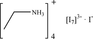
In the present article, we report the synthesis and crystal structure analysis of a new polyiodide compound, 4(C2H5NH3)+·I73–·I−.
2. Structural commentary
The title compound crystallizes in the monoclinic C2/m, with two ethylammonium cations and five I atoms in the asymmetric unit. In the extended structure, isolated I− and polyiodide [I7]3– anions are present. The positional disorder (Fig. 1) for the two cations is caused by the imposed mirror symmetry and that of the isolated anion (I5) by its special position (2/m; multiplicity 2, Wyckoff letter a).
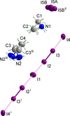 | Figure 1 The principal structural units in the crystal structure of the title compound. Displacement ellipsoids are drawn at the 50% probability level; short I—I bonds are drawn as dark solid lines, longer I—I bonds as bright solid lines. [Symmetry codes: (i) 2 − x, y, 1 − z; (ii) 1 − x, 1 − y, 2 − z; (iii) x, 1 − y, z.] |
Selected I—I bond lengths and angles of the [I7]3– polyanion are summarized in Table 1. The polyiodide can be described as two neutral I2 molecules (I2, I3) acting as connectors between three I− anions (I1, I4 and its symmetry-related counterpart) in a linear manner. The special position (2 c) of the central I1 atom results in point group symmetry 2/m for the entire polyanion. The I2—I3 distance is slightly longer than that in an elemental iodine molecule (2.72 Å; Svensson & Kloo, 2003). This is due to the n→σ* character of the halogen bond, which typically elongates the covalent bond of an I2 molecule. The distances between these I2 molecules and the three iodide anions (I1—I2 and I3—I4) are significantly longer. For comparison, similar I—I distances [2.8127 (5), 3.4270 (6) and 3.3875 (7) Å] are observed in [HMTAHD]3+[I7]3– (HMTAHD = 2,2,9,9,16,16-hexamethyl-2,9,16-triazoniaheptadecane; Lin et al., 2012). As seen in Table 1, the angles between the I atoms in the [I7]3– anion are all close to 180°. This polyiodide species adopts a unique linear conformation that has not been previously reported.
| ||||||||||||||||||||||
The negative charge of I− and [I7]3– anions is balanced by four ethylammonium cations. The C—C bond lengths are 1.522 (9) Å and 1.525 (11) Å and the C—N bond lengths are 1.478 (6) and 1.423 (10) Å, which are within the expected ranges for ethylammonium cations (Allen et al., 1987).
3. Supramolecular features
The shortest I⋯I distances between the isolated I− and [I7]3– anions are 4.5891 (5) Å [I5A⋯I2i; symmetry code: (i) − + x, − + y, z], 4.6691 (3) Å (I5A⋯I1i) and 5.3798 (4) Å (I5A⋯I4), which significantly exceed the sum of the van der Waals radii of 4.3 Å for two iodine atoms (Svensson & Kloo, 2003), indicating no significant covalent or non-covalent interactions between them. The layers of polyiodide anions exhibit a slipped-stack arrangement along the c axis (Fig. 2). The organic cations are located between the anionic chains and are linked to them via N—H⋯I hydrogen bonds (Fig. 3, Table 2).
|
 ; (ii)
 ; (iii)
 ; (iv)
 ; (v)
 .
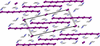 | Figure 2 The crystal structure of the title compound in a view approximately along the b axis. |
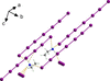 | Figure 3 A fragment of crystal structure showing the N—H⋯I hydrogen bonds between ethylammonium cations and both iodide and heptaiodide anions (N1—C1—C2 cation left; N2–C3—C4 cation right). |
4. Database survey
A search in the Cambridge Crystal Database (CSD, version 5.43, last update May 2026; Groom et al., 2016) for heptaiodide moieties, including both [I7]− and [I7]3– anions, revealed 33 entries. Among these, only two structures contain an additional, isolated I− anion co-crystallized alongside the heptaiodide moiety (JAPRUO, Antoniadis et al., 2005; ONOCOM, Agarwal, 2021).
To highlight the structural novelty of the title compound, it is compared with the closest discrete heptaiodide analogues: hexakis(2-hydroxypyridine) bis(2-hydroxypyridinium) hemikis(iodide) sesquikis(heptaiodide) diiodine (JAPRUO; Antoniadis et al., 2005) and 1,3-dimethyl-1H-imidazol-3-ium heptaiodide (YUPXOY; Fei et al., 2015). It is also worth noting the already mentioned structure of [HMTAHD]3+[I7]3– (PEKKEX; Lin et al., 2012). The three compounds exhibit significant structural differences compared to the linear [I7]3– conformation of the title compound. In both JAPRUO and YUPXOY, the [I7]− anions adopt bent (zigzag or Z-type) shapes described as I2·I3−·I2 adducts featuring short I—I bonds typical for I2 subunits. The only other [I7]3– species, PEKKEX, shares the same I−·I2·I−·I2·I− sequence. However, this polyiodide anion shows a longer I2 of 2.8127 (5) Å and adopts a V-shaped arrangement with an I⋯I⋯I angle of 136.22 (9)°.
5. Synthesis and crystallization
Ethylamine (0.5 mmol) was mixed with iodine (1 mmol) in 3 ml of 55%wt HI. The mixture was stirred for 4 h and then left to stand for one month. Black crystals formed during this period. The crystals were separated and stored under Paratone oil until the diffraction experiment.
6. Refinement
Crystal data, data collection and structure details are summarized in Table 3. The I5 atom representing the isolated iodide anion is disordered over a special position with site symmetry 2/m and was refined with occupancies of 0.5 for I5A and 0.25 for I5B to ensure a total charge of −1. The ethylammonium group N1—C1—C2 lies in a mirror plane; the hydrogen atoms attached to N1 and C2 are disordered around this mirror plane and were refined with an occupancy of 0.5 each. In the ethylammonium group N2—C3—C4, the C4 atom lies on a mirror plane; atoms N2 and C3, as well as all H atoms bonded to them, are disordered about this mirror plane and thus were refined with an occupancy of 0.5. The H atoms were positioned geometrically and were allowed to ride on their parent atom with Uiso(H) = 1.5Ueq(C) for methyl H atoms, Uiso(H) = 1.2Ueq(C) for methylene H atoms, and Uiso(H) = 1.2Ueq(N) for ammonium H atoms.
|
Supporting information
CCDC reference: 2596264
contains datablock I. DOI: https://doi.org/10.1107/S2056989026010844/wm5816sup1.cif
Structure factors: contains datablock I. DOI: https://doi.org/10.1107/S2056989026010844/wm5816Isup2.hkl
| 4C2H8N+·I73−·I− | F(000) = 1064 |
| Mr = 1199.57 | Dx = 2.708 Mg m−3 |
| Monoclinic, C2/m | Mo Kα radiation, λ = 0.71073 Å |
| a = 23.0274 (12) Å | Cell parameters from 6780 reflections |
| b = 7.0212 (3) Å | θ = 2.6–29.5° |
| c = 9.3382 (5) Å | µ = 8.43 mm−1 |
| β = 102.984 (5)° | T = 293 K |
| V = 1471.19 (13) Å3 | Plate, clear light orange |
| Z = 2 | 0.32 × 0.11 × 0.06 mm |
| XtaLAB Synergy, Dualflex, HyPix diffractometer | 2106 independent reflections |
| Radiation source: micro-focus sealed X-ray tube, PhotonJet (Mo) X-ray Source | 1734 reflections with I > 2σ(I) |
| Mirror monochromator | Rint = 0.061 |
| Detector resolution: 10.0000 pixels mm-1 | θmax = 30.4°, θmin = 2.6° |
| ω scans | h = −32→30 |
| Absorption correction: analytical [CrysAlisPro (Rigaku OD, 2024) using a multifaceted crystal model based on expressions derived by Clark & Reid (1995)] | k = −9→9 |
| Tmin = 0.043, Tmax = 0.491 | l = −12→12 |
| 10365 measured reflections |
| Refinement on F2 | Hydrogen site location: mixed |
| Least-squares matrix: full | H-atom parameters constrained |
| R[F2 > 2σ(F2)] = 0.032 | w = 1/[σ2(Fo2) + (0.0507P)2] where P = (Fo2 + 2Fc2)/3 |
| wR(F2) = 0.089 | (Δ/σ)max = 0.001 |
| S = 1.04 | Δρmax = 0.87 e Å−3 |
| 2106 reflections | Δρmin = −1.42 e Å−3 |
| 78 parameters | Extinction correction: SHELXL (Sheldrick, 2015b), Fc*=kFc[1+0.001xFc2λ3/sin(2θ)]-1/4 |
| 0 restraints | Extinction coefficient: 0.0059 (3) |
| Primary atom site location: dual |
Geometry. All esds (except the esd in the dihedral angle between two l.s. planes) are estimated using the full covariance matrix. The cell esds are taken into account individually in the estimation of esds in distances, angles and torsion angles; correlations between esds in cell parameters are only used when they are defined by crystal symmetry. An approximate (isotropic) treatment of cell esds is used for estimating esds involving l.s. planes. |
| x | y | z | Uiso*/Ueq | Occ. (<1) | |
| I1 | 1.000000 | 1.000000 | 0.500000 | 0.06616 (18) | |
| I2 | 0.85877 (2) | 1.000000 | 0.56630 (4) | 0.05694 (16) | |
| I3 | 0.74738 (2) | 1.000000 | 0.63002 (4) | 0.05321 (15) | |
| I4 | 0.61406 (2) | 1.000000 | 0.72342 (5) | 0.05892 (16) | |
| I5A | 0.500000 | 0.500000 | 1.000000 | 0.094 (2) | 0.5 |
| I5B | 0.500000 | 0.4245 (9) | 1.000000 | 0.0607 (8) | 0.25 |
| N1 | 0.61795 (17) | 0.500000 | 0.8233 (5) | 0.0533 (10) | |
| H1A | 0.615158 | 0.612508 | 0.778332 | 0.064* | 0.5 |
| H1B | 0.585030 | 0.478650 | 0.855725 | 0.064* | 0.5 |
| H1C | 0.622214 | 0.408841 | 0.760206 | 0.064* | 0.5 |
| N2 | 0.9570 (4) | 0.5833 (12) | 0.6813 (9) | 0.100 (3) | 0.5 |
| H2D | 0.929722 | 0.579762 | 0.597037 | 0.121* | 0.5 |
| H2E | 0.992363 | 0.551554 | 0.665169 | 0.121* | 0.5 |
| H2F | 0.958778 | 0.700361 | 0.718616 | 0.121* | 0.5 |
| C1 | 0.6701 (2) | 0.500000 | 0.9487 (6) | 0.0639 (14) | |
| H1D | 0.668800 | 0.388199 | 1.008890 | 0.077* | 0.5 |
| H1E | 0.668800 | 0.611801 | 1.008890 | 0.077* | 0.5 |
| C2 | 0.7280 (3) | 0.500000 | 0.8956 (8) | 0.0710 (17) | |
| H2A | 0.725886 | 0.404839 | 0.820771 | 0.107* | 0.5 |
| H2B | 0.760713 | 0.472271 | 0.976485 | 0.107* | 0.5 |
| H2C | 0.733746 | 0.622890 | 0.855907 | 0.107* | 0.5 |
| C3 | 0.9410 (5) | 0.4524 (12) | 0.7824 (13) | 0.091 (3) | 0.5 |
| H3A | 0.969266 | 0.461066 | 0.876466 | 0.109* | 0.5 |
| H3B | 0.941948 | 0.323429 | 0.745838 | 0.109* | 0.5 |
| C4 | 0.8785 (4) | 0.500000 | 0.8011 (9) | 0.100 (3) | |
| H4A | 0.855137 | 0.548498 | 0.710193 | 0.150* | 0.5 |
| H4B | 0.881024 | 0.594414 | 0.876594 | 0.150* | 0.5 |
| H4C | 0.860145 | 0.387021 | 0.828282 | 0.150* | 0.5 |
| U11 | U22 | U33 | U12 | U13 | U23 | |
| I1 | 0.0375 (3) | 0.1007 (4) | 0.0587 (3) | 0.000 | 0.0076 (2) | 0.000 |
| I2 | 0.0554 (2) | 0.0580 (2) | 0.0585 (2) | 0.000 | 0.01484 (16) | 0.000 |
| I3 | 0.0537 (2) | 0.0511 (2) | 0.0544 (2) | 0.000 | 0.01120 (15) | 0.000 |
| I4 | 0.0477 (2) | 0.0455 (2) | 0.0861 (3) | 0.000 | 0.02051 (18) | 0.000 |
| I5A | 0.0704 (13) | 0.100 (5) | 0.125 (3) | 0.000 | 0.0488 (14) | 0.000 |
| I5B | 0.0613 (13) | 0.064 (3) | 0.0609 (14) | 0.000 | 0.0225 (9) | 0.000 |
| N1 | 0.045 (2) | 0.056 (2) | 0.059 (2) | 0.000 | 0.0106 (18) | 0.000 |
| N2 | 0.075 (5) | 0.138 (8) | 0.093 (6) | 0.018 (4) | 0.031 (4) | 0.039 (5) |
| C1 | 0.054 (3) | 0.077 (4) | 0.056 (3) | 0.000 | 0.004 (2) | 0.000 |
| C2 | 0.052 (3) | 0.069 (4) | 0.085 (4) | 0.000 | 0.000 (3) | 0.000 |
| C3 | 0.097 (7) | 0.064 (9) | 0.106 (7) | −0.006 (5) | 0.010 (6) | 0.006 (5) |
| C4 | 0.130 (8) | 0.095 (5) | 0.089 (5) | 0.000 | 0.054 (6) | 0.000 |
| I1—I2 | 3.4445 (4) | N2—C3 | 1.423 (10) |
| I2—I3 | 2.7604 (6) | C1—H1D | 0.9700 |
| I3—I4 | 3.3747 (5) | C1—H1E | 0.9700 |
| I5A—I5B | 0.530 (6) | C1—C2 | 1.522 (9) |
| I5A—I5Bi | 0.530 (6) | C2—H2A | 0.9600 |
| I5B—I5Bi | 1.061 (13) | C2—H2B | 0.9600 |
| N1—H1A | 0.8900 | C2—H2C | 0.9600 |
| N1—H1B | 0.8900 | C3—H3A | 0.9700 |
| N1—H1C | 0.8900 | C3—H3B | 0.9700 |
| N1—C1 | 1.478 (6) | C3—C4 | 1.525 (11) |
| N2—H2D | 0.8900 | C4—H4A | 0.9600 |
| N2—H2E | 0.8900 | C4—H4B | 0.9600 |
| N2—H2F | 0.8900 | C4—H4C | 0.9600 |
| I3—I2—I1 | 177.961 (16) | C1—C2—H2A | 109.5 |
| I2—I3—I4 | 177.540 (16) | C1—C2—H2B | 109.5 |
| I5B—I5A—I5Bi | 180.0 | C1—C2—H2C | 109.5 |
| I5A—I5B—I5Bi | 0.000 (2) | H2A—C2—H2B | 109.5 |
| H1A—N1—H1B | 109.5 | H2A—C2—H2C | 109.5 |
| H1A—N1—H1C | 109.5 | H2B—C2—H2C | 109.5 |
| H1B—N1—H1C | 109.5 | N2—C3—H3A | 109.9 |
| C1—N1—H1A | 109.5 | N2—C3—H3B | 109.9 |
| C1—N1—H1B | 109.5 | N2—C3—C4 | 109.0 (7) |
| C1—N1—H1C | 109.5 | H3A—C3—H3B | 108.3 |
| H2D—N2—H2E | 109.5 | C4—C3—H3A | 109.9 |
| H2D—N2—H2F | 109.5 | C4—C3—H3B | 109.9 |
| H2E—N2—H2F | 109.5 | C3ii—C4—C3 | 25.3 (6) |
| C3—N2—H2D | 109.5 | C3—C4—H4A | 109.5 |
| C3—N2—H2E | 109.5 | C3ii—C4—H4A | 100.3 |
| C3—N2—H2F | 109.5 | C3ii—C4—H4B | 91.8 |
| N1—C1—H1D | 109.5 | C3—C4—H4B | 109.5 |
| N1—C1—H1E | 109.5 | C3—C4—H4C | 109.5 |
| N1—C1—C2 | 110.9 (5) | C3ii—C4—H4C | 134.0 |
| H1D—C1—H1E | 108.0 | H4A—C4—H4B | 109.5 |
| C2—C1—H1D | 109.5 | H4A—C4—H4C | 109.5 |
| C2—C1—H1E | 109.5 | H4B—C4—H4C | 109.5 |
| N2—C3—C4—C3ii | −39.0 (8) |
| Symmetry codes: (i) −x+1, −y+1, −z+2; (ii) x, −y+1, z. |
| D—H···A | D—H | H···A | D···A | D—H···A |
| N1—H1A···I4 | 0.89 | 2.77 | 3.6283 (12) | 163 |
| N1—H1B···I5A | 0.89 | 2.62 | 3.481 (4) | 163 |
| N1—H1B···I5B | 0.89 | 2.64 | 3.521 (4) | 170 |
| N1—H1B···I5Bi | 0.89 | 2.70 | 3.521 (4) | 154 |
| N1—H1C···I2iii | 0.89 | 3.23 | 3.794 (4) | 123 |
| N1—H1C···I4iv | 0.89 | 2.89 | 3.6283 (12) | 141 |
| N2—H2D···I4iii | 0.89 | 2.99 | 3.811 (8) | 155 |
| N2—H2E···I4v | 0.89 | 2.76 | 3.597 (9) | 158 |
| N2—H2F···I5Bvi | 0.89 | 3.03 | 3.775 (9) | 143 |
| Symmetry codes: (i) −x+1, −y+1, −z+2; (iii) −x+3/2, y−1/2, −z+1; (iv) x, y−1, z; (v) x+1/2, y−1/2, z; (vi) x+1/2, y+1/2, z. |
| I—I bond | Length | I—I—I | Angle |
| I1—I2 | 3.4445 (4) | I1—I2—I3 | 177.961 (16) |
| I2—I3 | 2.7604 (6) | I2—I3—I4 | 177.540 (16) |
| I3—I4 | 3.3747 (5) | I2—I1—I2i | 180.0 |
| Symmetry code: (i) 2 - x, y, 1 - z. |
Acknowledgements
The authors are grateful to the FAIRE programme provided by the Cambridge Crystallographic Data Centre (CCDC) for the opportunity to use the Cambridge Structural Database (CSD) and associated software.
Funding information
Funding for this research was provided by: Ministry of Education and Science of Ukraine (grant No. 24BF037-02).
References
 Agarwal, R. A. (2021). Cryst. Growth Des. 21, 2046–2055. CrossRef CAS Google Scholar
Allen, F. H., Kennard, O., Watson, D. G., Brammer, L., Orpen, A. G. & Taylor, R. (1987). J. Chem. Soc. Perkin Trans. 2 pp. S1–18. Google Scholar
Antoniadis, C. D., Hadjikakou, S. K., Hadjiliadis, N., Kubicki, M. & Butler, I. S. (2005). New J. Chem. 29, 714–720. Web of Science CrossRef CAS Google Scholar
Clark, R. C. & Reid, J. S. (1995). Acta Cryst. A51, 887–897. CrossRef CAS Web of Science IUCr Journals Google Scholar
Dolomanov, O. V., Bourhis, L. J., Gildea, R. J., Howard, J. A. K. & Puschmann, H. (2009). J. Appl. Cryst. 42, 339–341. Web of Science CrossRef CAS IUCr Journals Google Scholar
Fei, Z., Bobbink, F. D., Păunescu, E., Scopelliti, R. & Dyson, P. J. (2015). Inorg. Chem. 54, 10504–10512. Web of Science CSD CrossRef CAS PubMed Google Scholar
Groom, C. R., Bruno, I. J., Lightfoot, M. P. & Ward, S. C. (2016). Acta Cryst. B72, 171–179. Web of Science CrossRef IUCr Journals Google Scholar
Küpper, F. C., Feiters, M. C., Olofsson, B., Kaiho, T., Yanagida, S., Zimmermann, M. B., Carpenter, L. J., Luther, G. W., Lu, Z., Jonsson, M. & Kloo, L. (2011). Angew. Chem. Int. Ed. 50, 11598–11620. Google Scholar
Lin, J., Martí-Rujas, J., Metrangolo, P., Pilati, T., Radice, S., Resnati, G. & Terraneo, G. (2012). Cryst. Growth Des. 12, 5757–5762. CrossRef CAS Google Scholar
Mooney, R. C. L. (1935). Z. Kristallogr. 90, 143–150. CrossRef CAS Google Scholar
Rigaku OD (2024). CrysAlis PRO. Rigaku Oxford Diffraction, Yarnton, England. Google Scholar
Sheldrick, G. M. (2015a). Acta Cryst. A71, 3–8. Web of Science CrossRef IUCr Journals Google Scholar
Sheldrick, G. M. (2015b). Acta Cryst. C71, 3–8. Web of Science CrossRef IUCr Journals Google Scholar
Sonnenberg, K., Mann, L., Redeker, F. A., Schmidt, B. & Riedel, S. (2020). Angew. Chem. Int. Ed. 59, 5464–5493. Web of Science CSD CrossRef CAS Google Scholar
Svensson, P. H. & Kloo, L. (2003). Chem. Rev. 103, 1649–1684. Web of Science CrossRef PubMed CAS Google Scholar
Westrip, S. P. (2010). J. Appl. Cryst. 43, 920–925. Web of Science CrossRef CAS IUCr Journals Google Scholar
This is an open-access article distributed under the terms of the Creative Commons Attribution (CC-BY) Licence, which permits unrestricted use, distribution, and reproduction in any medium, provided the original authors and source are cited.
| CRYSTALLOGRAPHIC COMMUNICATIONS |
access