

 journal menu


issue contents
August 2008 issue
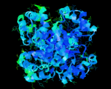
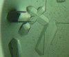
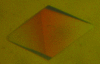
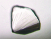
Cover illustration: Cytochrome c6 from the brown alga Hizikia fusiformis (Akazaki et al., p. 674).
protein structure communications


The crystal structure of cytochrome c6 from the brown alga H. fusiformis has been determined at 1.6 Å resolution. The amino-acid sequence and tertiary structure of H. fusiformis cytochrome c6 were very similar to those of red algal cytochrome c6 rather than those of green algal cytochrome c6.
PDB reference: cytochrome c6, 2zbo, r2zbosf
A combination of spectroscopic and crystallographic data have been used to identify the native metal-ion preference of GpdQ. These data suggest that GpdQ is a homobinuclear non-haem iron enzyme.
A crystal structure of zebrafish Plk1 reveals the binding of an extended ATP-competitive inhibitor that targets the adaptive pocket and helps to order the activation segment of the kinase. The conformation of the segment resembles that observed in a nonphosphorylated ligand-free wild-type kinase domain.
crystallization communications

The gene-regulation factor PyrR from B. halodurans has been crystallized in two crystal forms. Preliminary crystallographic analysis showed that the protein forms tetramers in both space groups.
Crystallization and preliminary X-ray analysis of L-methionine γ-lyase 1 from Entamoeba histolytica
L-Methionine γ-lyase 1, a key enzyme in sulfur-containing amino-acid degradation, from the protozoan parasite E. histolytica was crystallized in a form suitable for X-ray structure analysis.
The structure of the decameric inducible lysine decarboxylase from E. coli was determined by SIRAS using a hexatantalum dodecabromide (Ta6Br122+) derivative. Model building and refinement are under way.
Crystallization of the cystine-knot protein Spätzle occurred following serendipitous limited degradation of the pro-Spätzle propeptide during the crystallization experiment.
Crystals of the soluble ternary GM-CSF receptor complex were obtained which diffracted to a resolution of 3.3 Å.
The N-terminal moiety of C. thermocellum endo-1,4-β-D-xylanase 10B, comprising a carbohydrate-binding module (CBM22-1) and a GH10 E337A mutant domain, has been crystallized in complex with xylohexaose. The crystals belong to the trigonal space group P3221, contain a dimer in the asymmetric unit and diffract to beyond 2.0 Å resolution.
This article describes the first successful crystallization of a membrane-bound [NiFe] hydrogenase isolated from a photosynthetic organism (A. vinosum). The crystals obtained produced diffraction patterns up to 2.5 Å resolution.
Glyceraldehyde-3-phosphate dehydrogenase A has been cloned, expressed and purified. Apoprotein crystals have been grown which diffracted to 1.75 Å resolution and belonged to space group P21; holo crystals were grown in the presence of NADP, diffracted to 2.6 Å resolution and belonged to space group P32.
Glyceraldehyde-3-phosphate dehydrogenase B from H. pylori has been cloned, expressed, purified and crystallized in the presence of NAD. Crystals of GAPDHB diffracted to 2.8 Å resolution and belonged to space group P6522, with unit-cell parameters a = b = 166.1, c = 253.1 Å.

Preliminary X-ray data collection and analysis for crystals of chlorite dismutase, a haem-based enzyme that very effectively reduces chlorite to chloride while producing molecular oxygen, is reported to 2.1 Å resolution.
A 2S albumin from L. culinaris was purified and crystallized and preliminary crystallographic studies were carried out.
Maleylacetate reductase from Rhizobium sp. strain MTP-10005 has been crystallized using the sitting-drop vapour-diffusion method and microseeding. The crystals contained one dimeric molecule per asymmetric unit and diffracted to 1.79 Å resolution.

L-Asparaginase from H. pylori was overexpressed in E. coli, purified and crystallized. The crystals belonged to space group I222, with unit-cell parameters a = 63.6, b = 94.9, c = 100.2 Å and one molecule in the asymmetric unit. A complete data set to 1.6 Å resolution was collected using synchrotron radiation.
tRNA (m7G46) methyltransferase from E. coli was overexpressed, purified and crystallized. Diffraction data were collected to 2.04 Å resolution.

The ribonuclease HI domain of a bifunctional protein Rv2228c-CobC from M. tuberculosis has been crystallized as a fusion protein with maltose-binding protein in a form suitable for high-resolution crystallographic analysis.
XometC, a cystathionine γ-lyase-like protein from X. oryzae pv. oryzae and an antibacterial drug-target protein against bacterial blight, was cloned, purified and crystallized. Preliminary X-ray crystallographic analysis of XometC crystals was carried out.
The crystal of a 1,3-1,4-β-glucanase produced by Paecilomyces thermophila belongs to the hexagonal space group P6322, with unit-cell parameters a = b = 154.54, c = 87.62 Å.
Crystallization of recombinant IgG-binding protein expressed in Escherichia coli using the hanging-drop vapour-diffusion method is described. The crystals belonged to space group P212121, with unit-cell parameters a = 38.98, b = 43.94, c = 78.17 Å.

The enzyme mannosyl-3-phosphoglycerate synthase from R. xylanophilus has been expressed, purified and crystallized. The crystals belong to the hexagonal space group P6522 and diffract to 2.2 Å resolution.

The full-length LysR transcriptional regulator TsaR from C. testosteroni T-2 has been crystallized in two crystal forms and several native and derivative data sets have been collected using synchrotron and in-house X-ray sources.
addenda and errata
Free 
Free
![[publBio]](/logos/publbio.gif)


