

 journal menu


issue contents
March 2015 issue
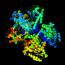
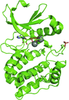
Cover illustration: Crystal structure of Escherichia coli tryptophanase (Kogan et al., p. 286).
IYCr crystallization series


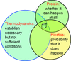
While crystallization phase diagrams are excellent tools to conceptualize phase relations in protein solutions, they need to be used appropriately and to be compatible with their physicochemical meaning.
research communications

Human myotubularin-related protein 1 (MTMR1) was crystallized using polyethylene glycol 20 000 as a precipitant. Diffraction data have been collected to 2.0 Å resolution using synchrotron X-rays.
Crystals of a complex between a fragment of TssM obtained by limited proteolysis and a nanobody have been obtained and diffracted to 1.9 Å resolution.


The purification, crystallization and molecular-replacement structure solution of two crystal forms of the β-catenin homolog HMP-2 from C. elegans are described.

The structure of the dodecamer of the aminopeptidase APDkam598 from the archaeon D. kamchatkensis was determined using X-ray diffraction data to a resolution of 3.0 Å and single-particle electron microscopy.
PDB reference: APDkam598, 4wwv
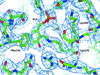
Two slightly different crystal forms of E. coli tryptophanase are presented. The transition between apo and holo forms of the enzyme is accompanied by a significant domain shift.

Cubic crystals of ArgR from B. halodurans have been obtained. Their diffraction was improved by the dehydration method. X-ray data have been collected to 2.35 Å resolution using synchrotron radiation.
Open  access
access
Two structures of Cwp84, a cysteine protease from the S-layer of C. difficile, are presented after propeptide cleavage. They reveal the movement of three loops, two in the active-site groove and one on the surface of the lectin-like domain, exposing a hydrophobic pocket.
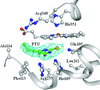
The crystal structure of the complex of lactoperoxidase with the antithyroid drug propylthiouracil (PTU) was determined at 2.50 Å resolution. It shows that PTU binds to lactoperoxidase in the substrate-binding site on the distal haem side. The S atom of PTU is coordinated to the haem iron, while the methyl group of the propyl moiety forms van der Waals contacts with the side chain of Ala114.
PDB reference: lactoperoxidase, complex with propylthiouracil, 4qyq
A carbohydrate-binding module from S. thermophile belonging to family GH64 was crystallized and diffracted to 1.8 Å. The phases were found using the single-wavelength anomalous diffraction method.
A variant of the Aurora A kinase domain in which both surface cysteines are mutated to alanine generates crystals that diffract to higher resolution than the equivalent crystals produced using wild-type protein. The structure confirms that single phosphorylation on Thr288 is insufficient to stabilize a conformation of the activation loop in a `fully active' state.
PDB reference: C290A:C393A Aurora A, 4ceg
Acetophenone reductase from G. candidium NBRC 4597 was crystallized by the hanging-drop vapour-diffusion method. The crystal diffracted to 2.6 Å resolution.
A protein construct consisting of a 149-residue fragment of human Caprin-1 was successfully expressed in E. coli, purified and crystallized. Native and Se-SAD data sets were collected to resolutions of 2.05 and 2.65 Å, respectively.

X-ray data were collected for two Dscam1 Ig7 isoforms and were processed to resolutions of 1.95 and 2.37 Å, respectively. Comparison of different Ig7 isoforms will provide insights into the mechanism of its homophilic binding specificity.
P. falciparum apicoplast DNA polymerase has been expressed, purified and crystallized in the apo form.
Open access
access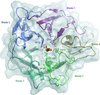
The crystal structure of the H. orenii glycosidase was determined by molecular replacement and refined at 1.10 Å resolution.
PDB reference: HoAraf43, 4qqs
Open access
access
The use of truncation and RNA-binding mutations of caffeine induced death suppressor protein 1 (Cid1) as a means to enhance crystallogenesis leading to an improvement of X-ray diffraction resolution by 1.5 Å is reported.
Rv1674c from M. tuberculosis, a possible DNA-binding sulfurtransferase, was purified and crystallized. Preliminary crystallographic analysis was performed.
![[publBio]](/logos/publbio.gif)


