

 journal menu


issue contents
October 2016 issue
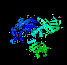
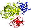
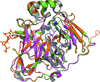
Cover illustration: The structure of glycoside hydrolase 29 family member GH29_0940, a protein cloned from metagenomic DNA from the rumen of a cow, reveals unique dual carbohydrate-binding domains (Summers et al., p. 750).
research communications


Open  access
access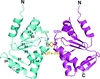
The dimeric structure of the C-terminal domain of Grx6, bridged by one [2Fe–2S] cluster coordinated by the active-site Cys136 and two external glutathione molecules, is reported.
The purification, crystallization and X-ray diffraction analysis of the Arabidopsis receptor-like cytoplasmic kinase BIK1, which plays an important role in growth and immune signaling pathways, are described.
Open access
access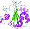
The crystal structure of mouse phospholipid hydroperoxide glutathione peroxidase 4 solved at 1.8 Å resolution and the first solution structural studies of a glutathione peroxidase protein are reported.
PDB reference: GPx4, 5l71
A high-resolution crystal structure of an unknown glycoside hydrolase enzyme from the rumen of B. taurus is presented. Unique dual carbohydrate-binding domains are revealed.
PDB reference: GH29_0940, 5k9h
Open access
access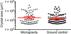
Microgravity was used in an attempt to define the crystal structure of the polyQ repeat of huntingtin alone or bound to MW1, an anti-polyQ Fab. While huntingtin was not crystallized in the experiments, analysis of microgravity-grown and Earth-grown MW1 Fab crystals showed that, on average, microgravity-grown crystals of MW1 Fab showed an increase in size and an improvement in resolution and mosaicity when compared with Earth-grown crystals in one space group, in agreement with data published for other proteins, although the highest overall resolution X-ray data in our experiments were obtained from a crystal grown on Earth.
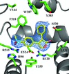
New crystallization conditions were identified that allowed the structure determination of human cyclooxygenase-2 in complex with rofecoxib (Vioxx) and the structure was subsequently determined to 2.7 Å resolution.
PDB reference: rofecoxib bound to human cyclooxygenase-2, 5kir
TtuA and TtuB are the sulfur transferase and sulfur carriers for the biosynthesis of 2-thioribothymidine in some bacterial tRNAs. To elucidate their mechanism of interaction, the TtuA–TtuB complex from T. thermophilus was crystallized and a Zn-MAD data set was collected to a resolution of 2.5 Å.
Open access
access
The extracellular domain of the A. thaliana ATP receptor DORN1 has been expressed and purified; a protein orthologous to DORN1 from C. sativa has been crystallized and its X-ray diffraction data have been collected.
Crystals of the multicopper oxidase CueO obtained from a perdeuterated sample for neutron diffraction diffracted to 1.8 Å resolution using X-rays and their crystal structure was solved.
The GDP-D-mannose pyrophosphorylase VTC1 from A. thaliana was crystallized and co-crystallized with its substrate. X-ray diffraction data were collected for crystallographic analysis.
![[publBio]](/logos/publbio.gif)


