journal menu


issue contents
January 2013 issue
Includes papers presented at the Seventh International Workshop on X-ray Damage to Biological Crystalline Samples
Diamond Light Source, UK, 14-16 March 2012
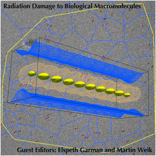
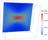
Cover illustration: Radiation damage montage: the background shows an overlay of the first and last image of an exposure series of cryo-electron microscopy images. The sample suffered from beam-induced motions; these motions were tracked using gold fiducial markers (in red). A Voronoi diagram, shown in blue, is used for non-rigid registration of the images (see Karimi Nejadasl, Karuppasamy, Newman, McGeehan and Ravelli, pages 58-66). In the foreground, absorbed dose isosurfaces (0.11, 1.8 and 2.5 MGy) for a cuboid crystal, exposed during an X-ray diffraction experiment ten times along its length using a translation data collection strategy to spread dose more evenly, as calculated by Zeldin, Gerstel and Garman (pages 49-57).
facility information


radiation damage


Free 
Introductory overview to the special issue papers on radiation damage to biological macromolecules.

The temperature and time dependence of global radiation damage are discussed in the context of outrunning damage at or near room temperature using synchrotron X-ray sources.
Open access
access
A systematic study of the sensitivity to radiation damage of crystals held at room temperature for a large set of model macromolecular structures is presented.
Open access
access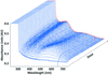
The reasons for the conflicting results on the efficacy of scavengers in macromolecular crystallography are examined in the light of some new results.
Singular value decomposition of a matrix is a versatile tool used in multivariate data analysis. Here, its use is presented to test the validity of physical models applied when scaling diffraction data affected by radiation-induced changes.

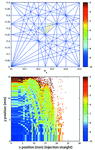
Simulated dose distributions for helical and translational macromolecular crystallography data-collection strategies are compared with that of a `standard' protocol as a step towards giving robust definitive guidelines to experimenters for optimizing the diffraction lifetime of crystals.
Open access
access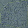
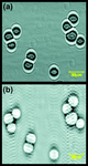
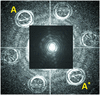
Cryo-electron microscopy images of vitrified large macromolecular complexes can become blurred due to beam-induced specimen alterations. Exposure series are examined, and rigid and non-rigid image registration schemes are applied to reduce such blurring.
research papers
Open access
access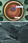
By using the strain-range partitioning method, the fatigue life of high-heat-load components made of oxygen-free copper have been successfully predicted within a factor of two.
Open access
access
Resonant inelastic X-ray scattering (RIXS) experiments require special sets of near-backscattering spherical diced analyzers and high-resolution monochromators for every distinct absorption-edge energy or emission line. For the purpose of aiding the design and planning of efficient RIXS experiments, a compilation of suitable crystal materials and viable reflections for hard X-rays, together with energy resolution and throughput information, is presented.
Open access
access
Using a convergent X-ray beam having continuously varying energy and glancing angle as a function of direction, the whole profile of a specular X-ray reflectivity curve is measured with no need for any mechanical motion during the measurement.
The optimal design of multilayer Laue lenses for a scanning X-ray microscope based on practical considerations of the apodization effect, monochromaticity requirement and working distance is presented.
An all-in-vacuum powder diffractometer yields the fundamental background level determined by Compton scattering and is ideal for high-resolution charge density measurements.
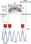
An original procedure, coupling synchrotron radiation with a mechanical device, is proposed in order to evaluate the strain-induced crystallization phenomenon at a crack tip in natural rubber during uninterrupted fatigue tests.
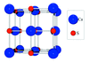
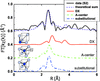
A discussion on the electronic structure and particular hybridization of CaS via ab initio calculations of X-ray absorption spectra is presented.
A statistical iterative image reconstruction algorithm is developed for the local tomography problem in synchrotron radiation X-ray micro-computed tomography.
An X-ray transmission cell was optimized for high-resolution in situ X-ray reflectivity measurements of the kinetics and thermodynamics of reactions at mineral–solution interfaces and its performance was tested using both optical and X-ray based measurements.
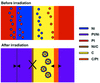
X-ray absorption spectroscopy, in conjunction with the X-ray standing-wave technique, is employed to characterize nanoclusters formed within a Pt/Ni/C multilayer by ion-irradiation.
A preliminary design of a knot undulator is presented. The characteristics of the radiation match the theoretical prediction well. The impact of the undulator on beam dynamics is benign.
A full-field X-ray imaging system based on the in-line germanium Bragg magnifier and Medipix detector is presented. A theoretical and experimental study of the imaging performance of the crystals–detector combination and a comparison with a standard indirect detector typically used in high-resolution X-ray imaging schemes are reported.
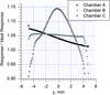
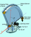
Measurements of the spatial variations in the response of three ionization chamber designs were tested as a function of chamber bias voltage, incident X-ray flux and fill gas.
EXAFS on Ga in CdTe complemented by DFT calculations with LAPW+lo methods provide evidence for DX- and A-centers as defect structures besides substitutional incorporation of Ga in CdTe〈Ga〉 grown crystals.
Open access
access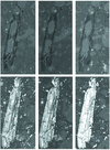
The 3D microstructure of shales is important to assess elastic anisotropic characteristics. In this study, microporosity and mineral components in two shale samples were investigated with X-ray tomographic microscopy at three synchrotron facilities: ALS, APS and SLS, and excellent agreement was observed.
A description of a new optical-transport-based set-up dedicated to soft X-ray magnetic resonant scattering in extreme sample environment, i.e. ultra-high vacuum, high magnetic fields and very low temperatures, is given.
short communications
It is shown that differently shaped prism arrays can provide spectral bandwidth for X-rays of the order of 0.5% under practically feasible experimental conditions.

A reaction furnace has been designed and built for use at the Materials Characterization by X-ray Diffraction beamline at the Elettra synchrotron source (Trieste, Italy). The furnace provides an atmosphere- and temperature-controlled environment for powders in capillaries and a temperature-controlled environment for thin-film samples.
The use of microbeams in X-ray absorption spectroscopy may lead to the oxidation of monomeric U(IV) species in environmental samples. Thus, care should be taken when analyzing samples that may contain monomeric U(IV) with X-ray microprobes.
computer programs
A program for deconvoluting multiple neighbouring absorption edges and computing values of f ′ and f ′′ is presented.
addenda and errata
Free

current events
Free