journal menu


issue contents
 | JOURNAL OF APPLIED CRYSTALLOGRAPHY |
High-resolution X-ray diffraction and imaging special issue (June 2017)
Guest Editors: Virginie Chamard and Václav Holý
The articles in this virtual special issue of Journal of Applied Crystallography represent some highlights of the 13th Biennial Conference on High-Resolution X-ray Diffraction and Imaging (XTOP 2016). The topics of the published articles cover the broad spectrum of problems discussed during the conference, highlighting in particular four specific X-ray techniques: X-ray Bragg diffraction, small-angle scattering and reflectivity, X-ray diffraction imaging, and coherent (phase-sensitive) X-ray imaging. These open-access articles were originally published in the journal between April and June 2017.
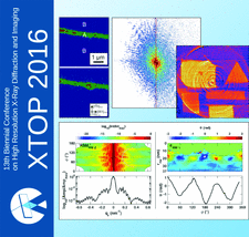
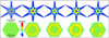
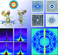
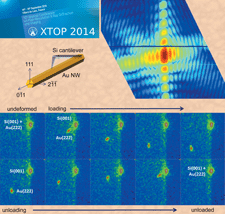
Cover illustration: Some highlights from XTOP 2016. Images courtesy of Schmidbauer et al. [J. Appl. Cryst. (2017), 50, 519–524], Hagemann & Salditt [J. Appl. Cryst. (2017), 50, 531–538], Jiang et al. [J. Appl. Cryst. (2017), 50, 712–721] and Davtyan et al. [J. Appl. Cryst. (2017), 50, 673–680].


 access
 access
 access
 access
 access
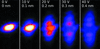 access
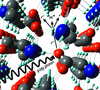
 access
 access
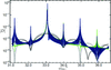 access
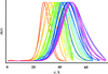
 access
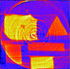 access
 access
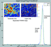 access
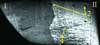 access
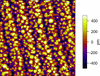 access
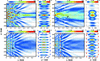 access
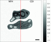 access