journal menu


issue contents
March 2009 issue
Includes papers presented at the Fifth International Workshop on X-ray Damage to Biological Crystalline Samples
Villigen, Switzerland, 3-5 March 2008
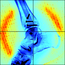
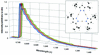
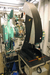
Cover illustration: Medical imaging and X-ray crystallography have one thing in common: striking a balance between data quality and radiation damage. James Holton (pictured) discusses how to best reach this compromise in X-ray crystallography (pages 133-142). He was not asked to discuss the same for medical imaging as his skills in this technique (as well as his skill at walking on icy pavements) were deemed questionable based on this image. The Swiss medical services near the Swiss Light Source are thanked for assistance in obtaining this image during the radiation damage meeting.
facility information


radiation damage


Free 

The current understanding of radiation damage in protein crystals is reviewed.
Open access
access
Radiation damage considerations affecting data collection by more than a factor of two are summarized and damage avoidance strategies are suggested.
Open access
access
Accurate measurement of photon flux from an X-ray source is a parameter required to calculate the dose absorbed by a sample. The development of a model for determining the photon flux incident on pin diodes, and experiments to test this model, are described for incident energies between 4 and 18 keV used in macromolecular crystallography.
The dose absorbed by a macromolecular crystal can be calculated using the program RADDOSE. A new version of this program, in which the probability of X-ray fluorescent escape following photoelectric absorption is included, is presented here with some examples of its application.
Open access
access
A portable and readily aligned online microspectrophotometer that can be easily installed on macromolecular crystallography beamlines is described. It allows measurement of the spectral characteristics of macromolecular crystals prior, during, and after the X-ray diffraction experiment.
Open access
access
Complementary techniques greatly aid the interpretation of macromolecule structures to yield functional information, and can also help to track radiation-induced changes. A new on-axis spectrometer being integrated into the macromolecular crystallography beamlines of the Swiss Light Source is presented.
Radiation-induced structural changes which can be explained by hydrogen abstraction were observed in a high-resolution X-ray diffraction study on cyclosporine A.
UV–Vis microspectrophotometry is used to test the efficacy of selected scavengers in reducing the undesirable photoreduction of the iron and copper centres in myoglobin and azurin, respectively, and X-ray crystallography to assess their capacity of mitigating global and specific radiation damage effects.
Two effective scavengers for room-temperature macromolecular crystallography are identified: ascorbate and 1,4-benzoquinone. Addition of these scavengers changes the usually observed diffraction intensity decay with dose from first order to zeroth order, leading to postulates on radiation damage mechanisms.
research papers
A `mini-beam' apparatus has been developed that conditions the size of an X-ray beam to 5 µm. The design of the apparatus and the characterization of the focal size and flux are presented.
A RATIO method for analysis of intensity changes in time-resolved pump–probe Laue diffraction experiments is described.
A two-dimensional photon-counting detector based on a micro-pixel gas chamber has demonstrated a dynamic range of >105 and a counting rate of up to 5 MHz.
Polycapillary-optics-based micro-XANES and micro-EXAFS at a third-generation bending-magnet beamline
A µ-XAS (µ-XANES and µ-EXAFS) set-up based on a polycapillary half-lens has been successfully tested at a bending-magnet station equipped with a non-fixed-exit monochromator.
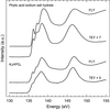
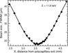
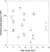
An overview of phosphorus L2,3-edge XANES spectra of pure reference compounds and geological phosphorus minerals, all measured under consistent experimental conditions, is given. In addition, the effect of radiation damage is shown for three compounds and measures are proposed to reduce it.
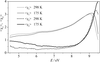
Valence electrons excitations of (NH4)2SO4 crystals have been studied by the spectral ellipsometry method using synchrotron radiation in the photon energy range E < 9.5 eV. Large anomalies of the temperature dependences of dielectric permittivity and intensity of reflected radiation have been found in the temperature range close to the phase transition point between para- and ferroelectric phases.
The instrumentation of the materials science X-ray beamline BL8 at the DELTA storage ring is presented together with its specific features. The first user experiments are also briefly described.
A new one-dimensional curved image-plate detector for rapid high-resolution powder X-ray diffraction investigations using synchrotron radiation is presented.
A bent-crystal spectrometer has been developed and experiments have been carried out in order to demonstrate the interests of a high-resolution detection system on a classical X-ray absorption beamline.

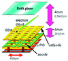
The results of X-ray absorption fine-structure experiments which were performed to test the applicability of a monolithic seven-cell silicon drift detector module developed at the Hamburger Synchrotron Strahlungslabor (DESY) for this type of measurement are described.
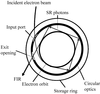
The performance of the far-infrared beamline of the 6 MeV MIRRORCLE-type tabletop synchrotron light source dedicated to far-infrared spectroscopy is presented.
short communications
Shear-induced rearrangement of colloidal aggregates was successfully imaged over time by X-ray microscopy, using a home-developed shear cell inserted into the focus plane. That experimental stimulation devices can be designed to meet the accuracy of X-ray microscopy extends the scope of its applications to dynamical processes.


This papers describes the study of a medieval prayer nut using synchrotron-based X-ray computed tomography, which revealed the fabrication method of the object.
current events
Free