journal menu


issue contents
January 2017 issue
Includes papers presented at the 9th International Workshop on X-ray Radiation Damage to Biological Crystalline Samples
Lund, Sweden, 9–11 March 2016
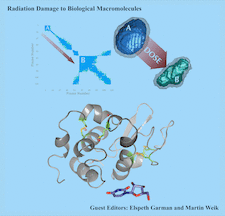
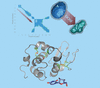
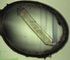
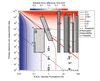
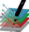
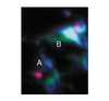
Cover illustration: (Designed by Charles S. Bury.) Top left: heatmap of results from pairwise comparison of the 1D scattering curves from a SAXS experiment on glucose isomerase (GI). Blue represents frames that show statistically strong pairwise similarity, whereas sea green shows weak pairwise similarity. Region A shows a cluster of frames exhibiting transitive pairwise similarity which were taken near the beginning of the experiment at low dose. Region B shows a large cluster of frames also exhibiting transitive pairwise similarity but which were generated when the sample had received a higher dose (see Brooks-Bartlett et al., pages 63–72). Top right: A – SAXS envelope generated using frames 1 to 11 (region A in left figure), exhibiting the expected globular shape of GI (MW = 173.85 kDa). B – SAXS envelope generated using frames 63 to 73 (high dose region B in left figure). The envelope does not exhibit the expected shape and corresponds to a much smaller molecular weight (MW = 173.85 kDa). The damage phenomenon causing this is predicted to be fragmentation. Bottom: Structure of uridine, a novel scavenger for MX and SAXS experiments, and the molecular structure of lysozyme at room temperature. Uridine increases the critical dose of lysozyme by 70% in MX experiments at room temperature, and shows scavenging properties in SAXS experiments at 40 mM concentration (see Crosas et al., pages pages 53–62).
facility information


radiation damage


 access
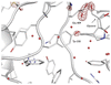 access
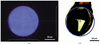 access
 access
 access
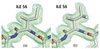 access
Bragg coherent diffraction imaging and metrics for radiation damage in protein micro-crystallography
research papers
Generation of apodized X-ray illumination and its application to scanning and diffraction microscopy
access access
 access
 access
 access
 access
 access
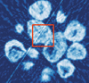 access
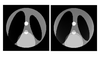 access
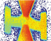

 access
beamlines
access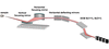
 access
 access
 access
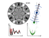
computer programs
current events