

 journal menu


issue contents
January 2020 issue
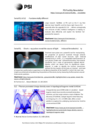
Cover illustration: RXES plane and extracted HERFD-XANES spectrum of IrO2, as an example of data obtained using the von Hamos-type hard X-ray spectrometer at the PETRA III beamline P64 (see Kalinko, Caliebe, Schoch and Bauer, pages 31-36).
facility information


research papers


Cinema:Bandit and its implementation in a continuous workflow for analyzing shock physics experiments and visually exploring the data in real time at X-ray light sources is presented.
Open  access
access
A THz plasma switch, driven by ultrafast XUV pulses for temporal ovelap in pump–probe experiments, is presented.

Single-particle diffraction is introduced as a simple but versatile technique to characterize the intrinsic properties of individual XFEL pulses such as total pulse energy and coherence.
It is shown that the high groove density and the large blaze angle of a recently successfully tested blazed grating for a spectrometer for space science are an optimum parameter set for operation in in-blaze off-plane diffraction in a monochromator for synchrotron radiation for the EUV/soft X-ray range. The instrument would provide competitive spectral resolution and superior transmission in comparison with use in in-plane diffraction.
Open access
access
The design and performance of the high-resolution wavelength-dispersive multi-crystal von Hamos type spectrometer at PETRA III beamline P64 are described.
Open access
access
White beam diagnostics through pinhole imaging of diffusely scattered radiation are presented.
A comparison of different material quality grades for coherent X-ray applications and the influence of the beryllium microstructure on the imaging and optical properties of compound refractive lenses are presented.
Open access
access
A new setup for picosecond pump–probe X-ray scattering at the Austrian SAXS beamline at Elettra-Sincrotrone Trieste is presented. Details of the specific implementation are discussed and results of a first transient-heating experiment are shown.
Open access
access
A new Rococo 2 X-ray fluorescence detector was implemented into the cryogenic sample environment at the Hard X-ray Micro/Nano-Probe beamline P06 at PETRA III, DESY, Hamburg, Germany. It features a high solid angle of up to 1.10 steradian and high count rates of 1 Mcounts s−1 per sensor.
A single-beam optical-trap sample holder for X-ray diffraction measurement of a single nanometre-sized particle was developed. The measurement for a single particle makes it possible to investigate the one-to-one relation between crystal structure and crystallite size of a particle.
The complex refractive index of beryllium was experimentally determined in the spectral range 20.4–250 eV from transmission spectra of free-standing thin films. A comparison of the optical constants of beryllium found in this way with those obtained earlier by another research group is performed.
Surface polarity has been demonstrated as a crucial parameter in determining physical properties and thus is of prime importance to electronic device applications in many transition metal oxide and semiconductor compound systems. This work presents the electronic structure variations of polar and nonpolar ZnO lattices with nitrogen-ion implantation using synchrotron-based in situ photoemission and X-ray absorption spectroscopy.
ZnO nanofoams synthesized using a soft template for solar photocatalysis including bacteria inhibition, and soft X-ray absorption spectroscopy studies used to study its morphology and doping are presented.
X-ray absorption near-edge structure (XANES) spectroscopy has been used to examine forms of aluminium (Al) in plants, with K-edge XANES suitable for identifying tetrahedrally and octahedrally coordinated Al. However, the poorer detection limit of L-edge XANES limits its use for plant tissues but, nevertheless, XANES analyses of Al in plants are of value where Al forms differ markedly.
Open access
access
Full-field spectroscopic measurements of the X-ray beam from a multilayer monochromator are presented. With these measurements, the striping artefacts often found when using these devices can be fully characterized.
Open access
access
A description and simulation of a full-field coherent imaging approach suitable for hard X-rays based on a classical (i.e. Galilean) X-ray microscope.


Using synchrotron radiation nano-computed tomography, the three-dimensional structures of micro-voids in 1,3,5-triamino-2,4,6-trinitrobenzene crystals were obtained with high spatial resolution and excellent imaging contrast, and then the development of the voids was followed in situ under different compressive strains.
Monte Carlo ray tracing is successfully used to simulate coherent diffractive imaging with a Huygens' wavelets-based approach.
The influence of grain size, coherence length and detector pixel size on the speckle-based X-ray imaging technique is discussed.
Phase-contrast X-ray tomography together with advanced image processing has provided 3D observation of unstained HeLa cells grown in PLLA electrospun scaffolds with a large field of view. The 4 µm fiber diameter scaffold showed cell clustering and deeper penetration after 8 days of growth compared with the 2 µm fiber diameter scaffold.
Open access
access
The imaging of live animals at a synchrotron source presents challenges in terms of remote monitoring and intervention, in addition to a sample that is changing and moving with time. This work describes experimental techniques that have been developed to address these challenges and capture image sequences that can equip a range of biomedical studies.
A correlative method based on fiducial markers visible in both Cryo-SXT and Cryo-FM was used to fuse images of two modalities precisely, and vacuoles and mitochondria were identified by this method.
Single cells of human fibroblasts with the iron-storage disease Friedreich's ataxia were analyzed with the ESRF's powerful ID16A `nano-imaging' beamline located in Grenoble, France. Unsurpassed absolute limits of detection were obtained, among which 180 iron atoms within an X-ray beam footprint of 30 nm × 50 nm under scanning mode conditions, providing a unique insight in the cell's iron architecture at the subcellular and organelle level. Seen from a broader perspective, brilliant synchrotron light can as such lift the veil on the amount and complex arrangement of metals at the subcellular scale.
This study explores the application of synchrotron radiation and conventional microcomputed tomography in evaluating bone-biopsy specimens. The study showed that all the represented elements of grafted sites and clear and accurate segmentations resulted in a realistic and detailed 3D image of woven bone structures such as new bone and bone substitutes.
short communications
Iterative energy self-calibration of Fe X-ray absorption near-edge structure data using the combination of two measures of Fe oxidation state from different parts of the spectrum.

Download citation


Download citation
Crystal structure of propionitrile (CH3CH2CN) determined using synchrotron powder X-ray diffraction
The structure and thermal expansion of propionitrile have been determined from 100 to 150 K using synchrotron powder X-ray diffraction.
CCDC reference: 1967900
beamlines
Time-resolved X-ray excited optical luminescence (XEOL) capabilities were developed successfully for the 23A X-ray nanoprobe beamline at the Taiwan Photon Source, with an excellent spatial resolution of <60 nm. The XEOL spectrum and decay time were simultaneously recorded over three time intervals, including normal (30 ps to 2 ns), hybrid (30 ps to 310 ns) and single (30 ps to 1.72 µs) bunch modes.
Download citation
Download citation
The experimental setup of the Xpress beamline at the Elettra synchrotron (Trieste, Italy) is described, along with the procedures for single-crystal centring and X-ray diffraction data collection and processing. The reported results demonstrate the possibility to perform in situ high-pressure single-crystal X-ray diffraction experiments at the Xpress beamline.
CCDC reference: 1965027
The microfluidic laboratory recently installed at Synchrotron SOLEIL is described. Three application examples are presented that showcase the opportunities offered by this facility to couple microfluidic systems with synchrotron techniques.
Open access
access
The performance of the recently commissioned spectrometer PEAXIS for resonant inelastic soft X-ray scattering and X-ray photoelectron spectroscopy and its hosting beamline U41-PEAXIS at the BESSY II synchrotron are experimentally characterized. With the design of the instrument, a 5 m-long RIXS spectrometer arm that can be continuously rotated by 106° and its sample environment for single crystalline materials covering a broad temperature range 10 K–1000 K, PEAXIS targets fundamental science in solid state physics where access to a range of wavevector transfers is imperative.


